Study of Hepatopancreas Tissue Damage in Mangrove Areas to Mud Crab (Scylla sp) Bombana Regency, Southeast Sulawesi, Indonesia
It is suspected that some of the mangrove near mining areas have a decline in quality due to mining activities. This study aims to determine the damage to hepatopancreas tissue in mud crab Scylla sp caught in the mangrove area in Bombana Regency, Southeast Sulawesi, Indonesia. The number of crab samples examined was 75 individuals with three sampling times with an interval of 1 month from July to September 2019. Histological examination was carried out on the hepatopancreas using the procedure for making histological preparations. The results of histological examination of the hepatopancreas found bacterial colonies, damaged epithelial desquamation, and necrosis. Tissue damages and Hg-absorption are thought to be due to changes in the environment where samples were taken in the mangrove area.
Introduction
Scylla sp is a species in the crustacean family that has a high economic value. The distribution and life cycle of mud crabs can be found in estuary areas, mangrove forest areas and in offshore areas that have muddy bottom substrates [1]. The mangrove ecosystem as a resource for coastal areas is a combination of physical and biological aspects known as ecological functions. The ecological functions of mangrove ecosystems include being a habitat (place to live), a place to find food (feeding ground), a place for care and rearing (nursery ground), and a spawning ground (spawning ground) for biotics [2]. Mud or mangrove crabs reared in mangrove vegetation areas have greater growth when compared to those reared in waters without mangroves, this proves that mangroves support the life of Scylla sp, whether it is providing food or nursery [3].
Mangrove crabs are often found on the coasts of Bombana, Central Buton, West Muna and North Konawe, Southeast Sulawesi in Indonesia. The mangrove vegetation in Bombana Regency grows relatively homogeneously and is in accordance with the environmental conditions of the mangrove crabs [4], which are utilized by the surrounding community. However, in several areas of this mangrove forest there has been a lot of damage and is in a state of threat of degradation. One of the causes of damage to mangrove forests is due to the entry of heavy metal waste into the waters due to mining activities. In addition to causing physiological changes in mud crabs, damage to the mangrove environment can cause histological changes in mud crabs, especially on very sensitive organs to physiological and environmental changes.
The hepatopancreas is a digestive organ in the crustacean class including mud crabs which also has important functions, including the absorption function which is characterized by the presence of microvilli cells which show a function of absorption, enzyme secretion [5], nutrient storage, metabolism, site of synthesis of vitellogenin other than the ovary, as well as detoxification [6]. As the main organ of xenobiotic detoxification in the crustacean class, the hepatopancreas is an organ that is very sensitive to physiological changes and environmental influences [7].
The changes that occur in the aquatic environment directly and indirectly maybe affect the structure and function of the hepatopancreas of crabs. Therefore, this research will confirm this conjecture.
Materials and Methods
Sampling
Sampling was carried out in the mangrove area in Bombana Regency (4°37’46.064”S, 122°0’46.856”E) and in the mangrove area in Lakara Village, Konawe Selatan district (4°28’3.372”S, 122°20’36.297” E) as a comparison. Catching mud crabs using traps measuring 40 x 25 x 20 cm, trap operations are carried out in mangrove waters at a depth of about 1-4 meters.
Furthermore, soaking the traps (soaking) was carried out between 5-9 hours during morning arrests and for afternoon arrests for 5-12 hours. The samples used were mud crabs weighing around 100-250 g total 40 individuals. Sampling was carried out 4 times with the number of samples for each collection, namely a total of 10 individuals, at intervals of 1 week per sampling (for 1 month). Samples of mangrove crabs taken from the mangrove area were then transported to Kendari alive.
Mercury Measurement
Brackish water and sediment in the area around crab fishing grounds were sampled for measurement of mercury content, according to the method described by Nur I, et al. [4]. Polyvinyl chloride (PVC) pipe was used to remove sediments from the sampling site down to a depth of around 20 cm. Prior to Hg measurement, the sediment samples were transported, sealed, and stored at 4°C using the same cold vapor atomic absorption spectrophotometry method (U.S. EPA method 245.1).
Histopathological Examination
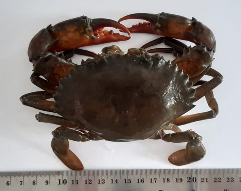
After taking samples of mangrove crabs first weighed to determine the weight, and introduction of the morphology of the mangrove crabs, then the mangrove crab samples were dissected using a knife and surgical scissors, then hepatopancreas samples were taken and put in onto the sample paper provided. Test animals are treated by complying with the code of ethics in animal testing.
The crabs are washed thoroughly under running water and then the hepatopancreas organs are taken using a scalpel and tweezers. Hepatopancreas organs were removed and fixed in 10% formaldehyde solution for 24 hours then histological preparations were made after going through the process of dehydration, clearing, embedding in paraffin, sectioning and stained with hematoxylin and eosin staining.
Data Analysis
The level of tissue damage in crabs was analyzed descriptively to determine the condition of the crab’s organ tissues
Results
Histology of Hepatopancreas Tissue Damage
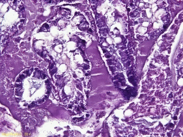
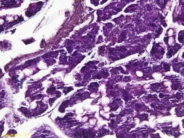
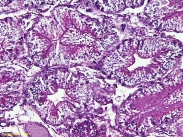
The figure below shows tissue damage to crabs caught in the mangrove area with mercury content in the sediment as shown in Figures 2 & 3.
A1
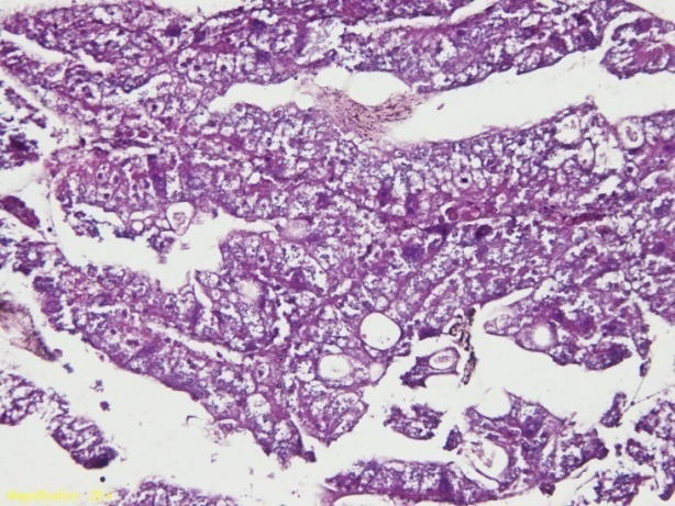
B2 Figure 2: Histology of Mud Crab Hepatopancreas. Magnification 200x. Severe heavy damages were seen in the Bombana Mud Crab samples (Figures A1 and A2) and light damage in the Mangrove Crab samples in Lakara Village (Figures B1 and B2). Hepatopancreas have necrosis tissues. (1) Liver cell necrosis, (2) Liver cell vacuolation (A1); (1) Liver cell necrosis, (2) Liver cell vacuolation, (3) Hemorrhagic (A2); (1) Vacuolization of liver cells, (2) Infiltration of mononuclear cells (B1); (1) Necrosis of liver epithelial cells (B2).
A2
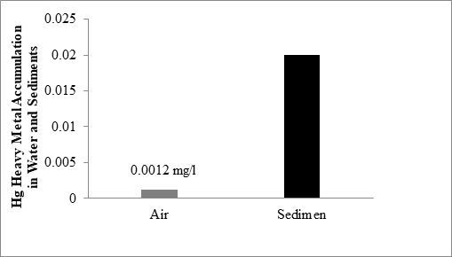
Mercury Content in Water and Sediments
The graph below shows the concentration of mercury content in water and sediments.
B1
Discussion
The hepatopancreas is the most important organ in crustaceans, because it functions as a digestive system, enzyme secretion, and nutrient storage. This is in accordance with the statement of Wang W, et al. [8] that the hepatopancreas is the most important organ in organisms, because this organ functions like the liver and pancreas in crustaceans. This organ produces digestive enzymes, stores nutrients, and removes waste.
Histopathological observations of the hepatopancreas of mud crabs in the Bombana area showed that there was damage in the form of hemorrhage, necrosis and severe vacuolization. Meanwhile, mangrove crabs in the Lakara area also had tissue damage in the form of necrosis and mild vacuolization (Figure 2). The changes occur because the cell nuclei and tubules in the hepatopancreas cause the shape between the tissues to become abnormal. This is in accordance with the statement of Bhavan PS, et al. [9] that, the hepatopancreas basically consists of branching tubules and various types of tubular epithelial layers, exposure to chemicals can cause structural changes in the tubular epithelial cells which cause histological changes. In addition, since the hepatopancreas is the primary organ for storage, metabolism, and detoxification, histopathological alterations in this organ may be caused by pesticide accumulation. Due to exposure to toxicant, it is possible that the crab’s tissue integrity was compromised as evidenced by the rupture of basal laminae in the hepatopancreatic tubules [10].
Hemorrhage is a condition characterized by bleeding from the vascular as a result of damage to the vascular wall. This is in accordance with the statement of Johnson AB, et al. [11] that, hemorrhage or bleeding is a condition characterized by the discharge of blood from the vascular as a result of damage to the vascular wall. The presence of hemorrhage can be caused by damage to the capillary endothelium due to infectious agents circulating in the blood vessels.
Necrosis is a change or death in cells. According to Takashima F, et al. [12] that, necrosis is tissue death resulting in tissue not being formed intact again. Necrosis of cells is usually caused by viruses, bacteria, fungi, and parasites or other chemical agents as well as disruption of the supply of blood to the body’s tissues resulting in shrinkage or reduction in the size of the nucleus as a whole. Cells that experience necrosis can be identified by the shape of their nuclei that shrink (pyknotic), enlarge, blur or disappear (karyolysis). Characteristics of necrotic tissue, which is paler than normal in color, loses vibrancy (tissue becomes brittle and tears easily), or has a poor or pale consistency. Cell necrosis is an uncontrolled cell death caused by biological agents such as viruses, bacteria, fungi, and parasites or chemical agents and interference with phagocyte antigens also decreases [13].
Vacuolization occurs due to tissue damage which is characterized by the loss of tubular epithelial cells that lose their cell contents or are empty. This is in accordance with the statement of Aliza D, et al. [14], that vacuolysis has characteristics such as round empty holes that occur due to fat accumulation. Factors causing vacuolysis are accumulation of toxic materials, lack of oxygen or excess fat consumption. If this vacuolysis does not disappear, it can interfere with the cell’s metabolic processes which are more severe and result in cell lysis. According to Nur I, et al. [15] that necrosis, hemorrhage and vacuolization of epithelial cells in the hepatopancreas and gills are the main histopathological changes due to disease.
Mononuclear cell infiltration is part of inflammation. This occurs due to interference with blood circulation. According to Suzuki Y, et al. [16], mononuclear cell infiltration or inflammatory cell infiltration is the entry of inflammatory cells into organ tissues in response to injury, toxic agents or disease.
Histological changes in aquatic organisms can provide information on susceptibility, stress levels, and adaptive abilities of organisms in dealing with stress. Histological changes can also be used as biomarkers and indicate a reaction to environmental changes. This is in accordance with the statement of Emiyarti E, et al. [17] that biomarkers in aquatic organisms can serve as early warning signals related to environmental threats posed and a useful tool for evaluating pollution loads in aquatic environments.
Conclusion
Samples of the hepatopancreas of mud crabs caught around the Bombana mangrove area have severe hemorrhage, heavy necrosis and vacuolization, whereas in the mangrove crab samples around the mangrove area of Lakara Village, South Konawe district, South East Sulawesi, Indonesia, there was only light tissue damages in the form of necrosis and mild vacuolization. This is due to the presence of heavy metal pollutants in the mangrove area.
Suggestions in this study are that it is necessary to carry out periodic environmental monitoring, especially in gold mining and there is a need for environmental improvement in the area.
Acknowledgments
Thanks to the staffs of Laboratory of Fisheries and Marine Science, Halu Oleo University and Laboratory of Pathology, Veterinary Research Centre, Bogor-Indonesia
for their technical assistance. We also want to thank the reviewers and journal editors for comments and suggestions to improve the manuscript for publication.
References
-
Muhtadi A, Yulianda F, Boer M, Kirisanti M (2022) Spatial distribution management of Crustacea (Decapoda) based on conservation in tropical tidal lake. Biodiversitas 23: 4064-4072.
-
Nagelkerken I, Blaber SJM, Bouillon S, Green P, Haywood M, et al. (2008) The Habitat Function of Mangroves for Terrestrial and Marine Fauna: A Review. Aquatic Botany 89(2): 155-185.
-
Putri A, Bengen DG, Zamani NP, Salma U, Kusuma NP, et al. (2022) Mangrove Habitat Structure of Mud Crabs Bee Jay Bakau Resort Probolinggo, Indonesia. Indonesian Journal of Marine Sciences 27(2): 124-132.
-
Nur I, Aris EA, Yusnaini Y, Beavis S (2021) The potential use of _Octolasmis_ spp. parasites in mud crabs _Scylla_ spp. as a bioindicator for mercury pollution. Biodiversitas 22(9): 3764-3772.
-
Zeng H, Haihui Y, Shaojing L, Guizhong W (2010) Hepatopancreas Cell Cultures from Mud Crab _Scylla_ _paramamosain_. in Vitro Cell Dev Biol Animal 46(5): 431- 437.
-
Sousa LG, Cuartas EI, Petriella AM (2005 Fine Structural Analysis of The Epithelial Cells in the Hepatopancreas of _Palaemonetes argentinus_ (Crustacea, Caridea). Biocell 29(1): 25-31.
-
Sousa LG, Petriella AM (2007) Functional Morphology of the Hepatopancreas of _Palaemonetes argentinus_ (Crustacea: Decapoda): Influence of Environmental Pollution. Int J Trop Biol 55(S1): 79-86.
-
Wang W, Wu X, Liu Z, Zheng H, Cheng Y (2014) Insights into hepatopancreatic functions for nutrition metabolism and ovarian development in the crab Portunus trituberculatus: gene discovery in the comparative transcriptome of different hepatopancreas stages. PLoS One 9(1): e84921.
-
Bhavan PS, Geraldine P (2009) Manifestation of Carbaryl Toxicity on Soluble Protein and Histopathology in the Hepatopancreas and Gills of the Prawn. _Macrobrachium_ _malcolmsonii_. Journal of Environmental Biology 30(4): 533-538.
-
Maharajan A, Narayanasamy Y, Ganapiriya V, Shanmugavel K (2015) Histological alterations of a combination of Chlorpyrifos and Cypermethrin (Nurocombi) insecticide in the fresh water crab, Paratelphusa jacquemontii (Rathbun), The Journal of Basic & Applied Zoology 72: 104-112.
-
Johnson AB, Hemorrhage BB (2023) In: StatPearls. Treasure Island (FL): StatPearls Publishing.
-
Takashima F, Hibiya T (1995) An Atlas of Fish Histology Normal and Phatology Feature. Tokyo Kodansha Ltd, pp: 108.
-
Khalid N, Azimpouran M, Necrosis (2023) In: StatPearls. Treasure Island (FL): StatPearls.
-
Aliza D, Nazaruddin, Abda AS, Asri A (2022) Mercury Chloride (HgCl2) Exposure Changes the Histopathological Figure of Eye and Brain of Tilapia Fish (_Oreochromis mossambicus_). Biotropia 29(2): 103-111.
-
Nur I, Yusnaini Y (2018) Parasites and Histopathology of Infected Spiny Lobster _Panulirus_ spp. Cultured in Outer of Kendari Bay, Indonesia. AACL Bioflux 11: 108-117.
-
Suzuki Y, Takaji I (1992) Fish granulocytes in the process of inflammation, Annual Review of Fish Diseases 2: 149- 160.
-
Emiyarti E, Nur I, Yusnaini Y, Astuti O, Patadjai RS (2020) Sublethal Toxicity Test of Mercury (Hg) in The Flesh and Tissue of Tilapia (_Oreochromis niloticus_). Omni-Akuatika 16(2): 99-107.
- California Red-Legged Frog and Non-Listed Amphibians Response to Non-Native Fish Removal
- Industrial Standardization of the Bio-OS: Algorithmic Codification of Resilience Engineering Guidelines and Version V8 Architecture
- Climate Variability and the Sustainability of Snail Farming in Nigeria: Past Trends, Present Challenges and Potential Outlook
- The Evaluation of the Surveillance System of Anthrax in Gilgit-Baltistan, Pakistan, 2018
- Natural Decline to Extinction of A New Zealand Rabbit Population
- Mitochondrial Bio-Logistics: Steering Co-Enzyme Q10 and Lycopene Synergies within the Science 4.0 Bio-OS Framework