Histopathological Investigation of Prawn Penaeus monodon in the Improved Extensive Culture Ponds of Nellore Region, Andhra Pradesh, India
Present work was aimed to see the quality of the prawn Penaeus monodon under improved extensive aquaculture system collected from Muthukur area of Andhra Pradesh located Pradesh located at 14.29 19 latitude and 80.10383 longitude. To assess the quality of the prawn, prawns were surveyed in the improved extensive pond and collected for histopathological investigation by adopting double staining methods (hematoxylin and eosin). Results showed that WSSV (White Spot Syndrome Virus) was characterised by prominent eosinophilic to pale basophilic intra nuclear inclusion bodies in hypertrophied nuclei of most commonly in the epithelial cells and connective tissue cells of the above target tissues. Monodon baculovirus (MBV) was characterised by intensive stained or multiple spherical or rounded hypertrophied intra nuclear inclusion bodies ranging from 0.1 m to 18 m in the epithelial cells of hepatopancreas. This kind of necrotic behaviour was also found along with acidophilic intra nuclear inclusion in hypertrophic nuclei with marginated chromatin. MBV infected shrimps showed generalised signs of disease such as lethargy, anorexia, poor feeding, dark colouration and reduced growth rate. Intensive stained basophilic hypertrophic rounded intranuclear inclusions symptoms were also found due to those disease both in eye and gill tissues. It is predicted that the prawn collected from the pond showed the symptoms of both viral and bacterial infection.
Introduction
The global aquaculture of penaeid shrimp has grown rapidly during the past two decades. In 1998 world shrimp farmers produced an estimated 737 200 t of whole shrimp (this includes 530 200 t from the eastern hemisphere and
207 000 t from the western hemisphere), a record 12% increase from 660 200 t produced in 1997 [1, 2]. The shrimp sector suffered a serious setback during 1995-96. Lack of technically qualified manpower, improper site selection, defective farm design, rapid intensification, overcrowding of farms in restricted locations and disproportionate development of the industry relative to supply of quality farm inputs paved the way for poor environmental conditions in ponds. The disease caused by a viral pathogen (white spot viral disease) during 1993-1994 delivered a lethal blow to the global shrimp farming industry, which could not recover immediately.
Shrimp diseases have caused many millions of dollars of losses over the past few years fueling research into their causes, diagnosis and the search to develop technologies to control and prevent them. Many such methods have been, and continued to be developed but their implementation at the farm level has in most cases been frustratingly slow. Transferring the benefits of new developments in shrimp health management and disease control into practical techniques for use in commercial farms in one of the biggest challenges facing shrimp aquaculturists. WSSV is considered the most serious problem for shrimp aquaculture in Asia. The virus affects all life stages of Penaeus monodon and mortality rate can reach 100% within 3-10 days of the onset of clinical signs Inouye, et al. [3]. These innovations have made shrimp culture more efficient in controlling diseases, more sustainable, and more environmentally friendly. Costa, et al. [4] reported Isolation and characterization of bacteria associated with a Penaeus stylirostris disease (syndrome 93) in New Caledonia and showed the presence of two different groups of vibrio sp. Populations of shrimp showing the signs of ‘White spot` disease display high mortality rates with cumulative mortalities reaching 100 within 3 to 10 days of the onset of clinical signs Momoyama, et al. [5]. White spot disease viruses are cylindrical to elliptical or obovate and measure 121+26nm in length Wongteerasupaya, et al. [6]. described the genome of the white spot disease virus as a double stranded DNA molecule longer than 150kbp [7, 8]. compiled the information pertaining to White spot causing viruses from different geographic regions along with the reported mean virion size and mean nucleocapsid size of these viral strains. White spot viral disease in penaeid shrimp is characterized histopathologically in most tissues of ectodermal and mesodermal origin [6]. MBV is known to infect Penaeus monodon, Penaeus merguensis, Penaeus semisulcatus and Penaues kerathurus Doubrovskyy, et al. [9]. MBV infection has also been reported in cultured Metapenaeus ensis in Taiwan Chen, et al. [10]. The natural reservoirs of MBV are believed to be wild Peneaus monodon and other susceptible species of shrimp [11]. Most earlier reports on the incidence of MBV studied in Asia has been based on histological data [12]. Pond-reared penaeid shrimp typically serve as hosts for a multitude of parasitic and epi commensal organisms Johnson, et al. [13] and their presence may not necessarily equate to disease. Gross and clinical signs, with the most commonly applied laboratory test being direct examination and microscopy using the light microscope, classical microbiology with isolation and culture of the agent, and routine histology and histochemistry [13].
‘Classic’ diagnostic techniques that are important, but are used less frequently, include techniques such as bioassay and enhancement, which are used for the detection of subclinical or carrier-state infections by certain pathogens Lightner, et al. [14]. Tapay, et al. [15] developed primers for PCR based on the sequence of a cloned fragment of the white spot disease virus genome and used the primers to detect white spot disease virus from both experimentally and naturally infected shrimp. Virucidal effects of ultraviolet (UV) radiation, heat, PH, Ozone, Salinity and some chemical disinfectants (sodium hypochlorite, povidone iodine and benzalkonium chloride) on White Spot disease virus were investigated by Chang, et al. [16] by infectivity assay using juvenile P. monodon. Sahul Hameed, et al. [17] reported the pathogenicity of systemic ectodermal and mesodermal baculo virus and its detection in shrimp by immunological methods. The result of their study showed the presence of SEMBV in all the organs and tissues expect in hepatopancreas. Eleonar, et al. [18] reported isolation of Vibrio spp. From Penaeus monodon (fabricius) with red disease syndrome and showed that red disease syndrome is characterized by the reddening of the shrimp body and isolated 4 Vibrio phenotypes namely Vibrio harveyi, Vibrio parahaemolyticus, Vibrio flurialis and Vibri sp. From shrimps with red disease. Indrani Karuna Sagar, et al. [19] reported that White Spot Syndrome Virus of Penaeus monodon along the west coast of India. Smith PT [20] reported toxic effects old blooms of marine species of Oscillatoriales on farmed prawns as (Penaeus monodon, Penaeus japonicus and brine shrimp Artemia salina secondary infection by pathogenic bacteria. Tomoya kono, et al. [21] studied on the detection on white spot syndrome virus in shrimp by loop mediated isothermal amplication and gave a standardized lamp procedure to detect the presence of WSSV in the heart, stomach and lymphoid organs from infected shrimp. Piamsak Menasveta [22] studied on Improved shrimp grow out systems for disease prevention and environmental sustainability in Asia and suggested that the most practical preventive measures is the improvement of grow out systems and showed the intensive system modified to a more biosecure system that is a closed recirculating water system, a reduced or zero water exchange system and shrimp culture at inland locations away from coastal influences. Hung-Hung sunga, et al. [23] studied on relationships between disease outbreak in cultured tiger shrimp (Penaeus monodon) and the composition of vibrio communities in pond water and shrimp hepatopancreas during cultivation and suggested that the presence of a large number of vibrio in the hepatopancreas may be associated with growth retardation in shrimps. Realizing the importance of the work, the present work was attempted on Penaeus monodon grown in the improved extensive culture pond which is located in Nellore region of Andhra Pradesh, India for the investigation of disease outbreak by conducting histological work.
Materials and Methods
The present study was carried out in Nellore region of Andhra Pradesh, India since the Nellore region is actively engaged with aquaculture practices on fish and prawn farming. With intensification of these activities in this area, the aquaculture sector has become benefited in one way and another way it is non-benefited because of outbreak of diseases in the cultivable species of fish and prawn with improper management of culture pond. The tiger prawn Penaeus monodon routinely cultured in this area have become pathogenic attack of virus like White Spot Syndrome Virus (WSSV), Monodon Bacculo Virus (MBV) and Hepatopancreatic Virus (HPV) etc. The details for pond management containing environmental parameters of water, water exchange, feed application, stocking density of the candidate species, mode of water supply etc. were collected and interpreted for the result of this study. To know the reason for causing of this disease in the candidate species of prawn, the present work has been attempted on histopathological investigation of prawn in Nellore region in relation with pond management. During the study period weekly samples of tiger prawn Penaeus monodon were collected from the improved extensive culture ponds for histopathological investigation, for which the tissues like hepatopancreas gills, eyestalk, soft tissue and gut were dissected out in live condition and immediately fixed in Davidson’s fixative for 24 hours. Tissues fixed in Davidson’s fixative were transferred to 70% ethanol after two days for further study. The target tissues like hepatopancreas, gills, eyestalk, soft tissue, fixed in Davidson’s fixative were subjected for histopathological work as given below.
Processing and Sectioning
The tissues of hepatopancreas, gills and eyestalk fixed were washed overnight in running tap water to remove the excess of picric acid. The fixed tissues were dehydrated using and alcohol series (30%-100%) and cleared in xylol. The tissues were further cold impregnated overnight with wax using xylene and wax shavings in 1:1 ratio. Subsequently the sample was evaporated by placing the tissues in an oven at 58 C. The tissues were then transferred through two changes of fresh molten wax (Paraffin wax M.P 58-60 C). The tissues embedded blocks were prepared by using proper orientation of wood or metal. Serial sections of the block were cut at approximately 6-8 m thickness using a rotary microtome. Sections were fixed on clear glass slides using fresh Mayer’s egg albumin and flattered by placing on a slide warmer with a drop of distilled water. Subsequently the water was drained off and the slides were allowed to dry. These slides were then used for histological staining.
Staining
Routine staining for gross morphological observations was carried out using Harris Hematoxylin stain with 1% alcoholic eosin as the counter stain. Sections to be stained were first deparaffinized in two changes of xylene and then hydrate through a descending series of propanol grades. Sections were blued using tap water or ammonia solution. Eosin stained sections were repeatedly washed in an ascending series of propanol grades to remove excess eosin and cleared in xylene. Sections were mounted with DPX mountant and examined under microscope.
Results
Since the collection spot of prawn Penaeus monodon under super extensive aquaculture system carried out in Muthukur area of Nellore region subjected under outbreak of disease, the present work was carried out to find out the type of diseases (bacterial and viral infection) happened in the particular pond ecosystem. During the present study, prawns were monitored for the occurring of diseases and along with this, certain water parameters were also monitored. Bore water containing 10-15 ppt saline was used both in extensive and improved extensive shrimp ponds for Penaeus monodon. Water was exchanged 10-25% irrespective of growth phase of prawn, the pH was ranged between 7.8-8.5 and turbidity became clear with slightly green in colour. Cp brand fed was routinely given in the culture pond starting from larvae to adult prawn in most of the farms studied. When the water become acidity in condition, the lime was added with quantity of 25kg/acre once in a week. 12,000-15,000 seeds/ acre were stocked in extensive pond and 25,000-30,000 seeds/acre were stocked in improved ponds. There was no scientific method of aeration provided in the culture ponds. Prawns were harvested at 30 counts /kg after the days from 110-120.
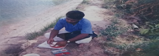
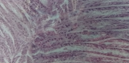
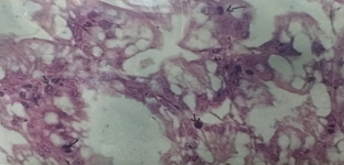
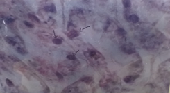
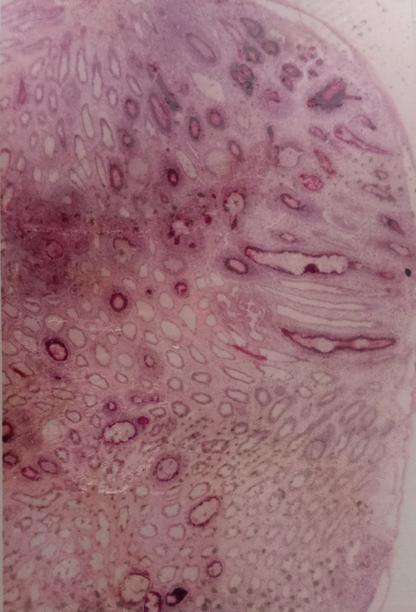
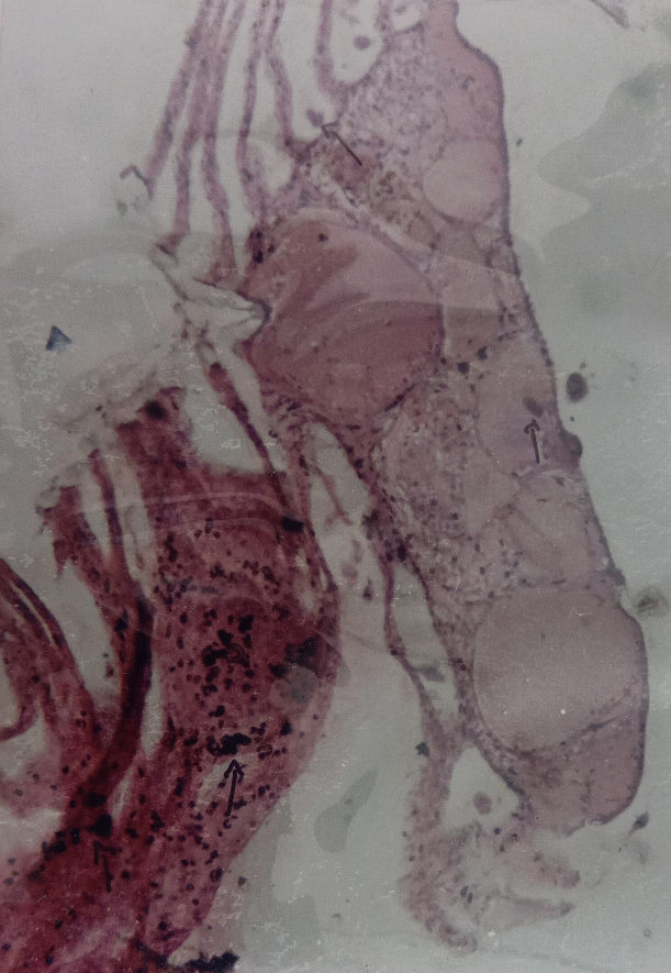
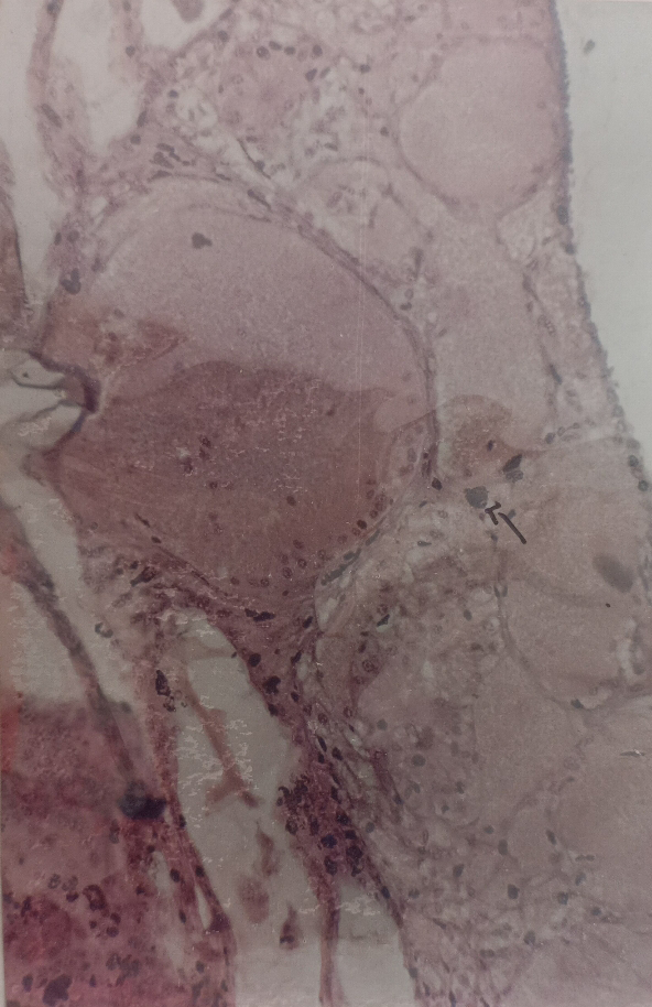
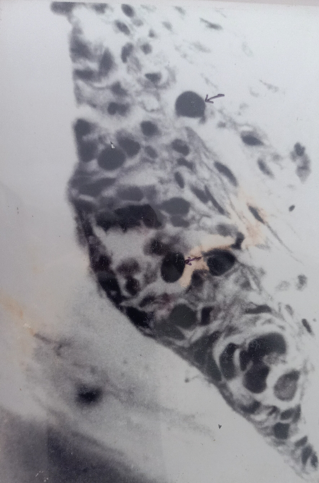
Target tissues such as gills, guts, eye tissue and soft tissues of cephalothoracioc region were dissected out from the infected animals of Penaeus monodon suspected with the disease of white spot syndrome virus (WSSV). WSSV was characterised by prominent eosinophilic to pale basophilic intranuclear inclusion bodies in hypertrophied nuclei of most commonly in the epithelial cells and connective tissue cells of the above target tissues. Expansion of the cuticular chromatophores was also noticed in the exoskeleton of Penaeus monodon. This causative syndrome might perhaps be due to the presence of WSSV. Further, it is suspected to be attacked with not only a single factor of virus but also with other target secondary bacterial infection (Figures 1-3). Characterisation of WSSV is shown in Figures 4-6 and collection and monitoring of the tiger prawns from the pond is shown in Figures 7-10 infected region of eye tissues is predicted in Figures 8 & 9. Monodon baculovirus (MBV) was characterised by intensive stained or multiple spherical or rounded hypertrophied intranuclear inclusion bodies. Result of the study showed that gill, hepatopancreas, eye and stomach are severely affected with the outbreak pathogens.
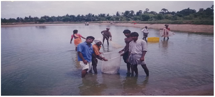
Figure1: Showing a Survey on Monitoring Shrimp Diseases in Penaeus monodon in the Culture Pond.

Discussion
Most of the economy of India from aquaculture sector depends on the euryhaline type of fin and shell fishes. Among the shell fishes, species of tiger prawn Penaeus monodon is placed in an important role for the culture value and export. Penaeid and non penaeid prawns have been affected due to improper management of pond, over stocking density and commercial feed containing antibacterial residues etc. Due to various factors, the disease outbreak has become quite common with various levels in the prawns in an unfavourable condition, particularly with WSSV, MBV, HPV and SEMBV etc. In the present study, WSSV, MBV and other bacterial diseases were noticed to cause the major damage or necrosis in the target tissues of Hepatopancreas, Gills, Gut, Eye stock and stomach, etc in the species of tiger prawn Penaeus monodon. Characterisations made at WSSV and MBV were quite common in the tissues for the present study as revealed by many authors in the same species Lightner, et al. [24] and it was prominently noticed when the study was made at Nellore area in improved extensive shrimp culture pond located at Muthukur area of Andhra Pradesh.
The results of this study showed that MBV is widely present in hatcheries in India. Presence of MBV need not necessarily result in disease and mortalities as this virus is well tolerated by Penaeus monodon in light to moderate infections [24]. It has been suggested that the transmission of MBV occurs through oral route in water contaminated with MBV from faecal matter of brood stock Chen, et al. [25]. It has also been proposed that MBV infection of eggs/larvae can be avoided by washing the fertilised eggs in clean filtered sea water. The difference observed in the incidence in different hatcheries may be due to variations in incidence of MBV in brood stock or due to hygienic practices in the hatchery. It has been suggested that MBV is well tolerated by Penaeus monodon in low to moderate infection if other conditions are optimal Lightner, et al. [24].
Nilskautsky, et al. [26] studied on ecosystem perspectives on management of disease in shrimp pond farming and discussed, that from an ecological perspective, the causes behind the development and spreading of pathogens in shrimp aquaculture, the risk of disease shrimp farming often increases with culture intensity and high stocking densities, and when polyculture is replaced by monoculture studied on detection of white spot baculovirus (WSBV) in giant freshwater prawn, Macrobrachium rosenbergii, using polymerase chain reaction and suggested that the amplified product from the DNA of the naturally- infected WSS Macrobrachium rosenbergii was similar to that of WSBV- infected Penaeus monodon. Rajan, et al. [27] studied on white spot baculovirus syndrome in the Indian shrimp Penaeus monodon and Penaeus indicus and suggested that infected Juveniles and sub adults of Penaeus indicus and Penaeus monodon become lethargic surface frequently, exhibit loss of difference with reduced feeding activities. It has been suggested that MBV is well tolerated by Penaeus monodon in low to moderate infection if other conditions are optimum. However, the production rate of the species concerned has become low [28, 29, 30, 31, 32, 33, 34, 35].
In the present study, WSSV was clearly seen in the gill of Penaeus monodon with its specific characteristic features. This disease was also found to be noticed in the target tissues of hepatopancreas, gills, gut etc [36, 37, 38, 39, 40, 41]. WSSV, MBV and other bacterial disease were also found to attack in Penaeus monodon mainly due to improper management of water, over stocking density, high feed dosage and failure of aeration. It is suggested from this study that the endemic species of Penaeus monodon and Penaeus indicus will have to be encouraged further for their higher production in extensive and semi intensive aquaculture system by promoting improvised clean pond ecosystem along with constant maintenance of feed quality which would be preferred by the species concerned [42, 43, 44, 45, 46, 47, 48, 49, 50, 51, 52, 53]. It is also suggested that the species Penaeus monodon is more compatible and candidate species than any other exotic species provided that the prawn should become more immunity against the attack from various kind of pathogens.
Acknowledgement
Authors are thankful to the authority of the Thiruvalluvar University, Vellore, Tamil nadu, India for providing necessary facilities.
References
-
Rosenberry B (1998) Shrimp hits new high. Fish farming International 25(12): 1.
-
Rosenberry B (1999) Farmed shrimp up by 12 percent. Fish farmer pp: 40-41.
-
Inouye K, Miwa S, Oseko N, Kimura T, Nakano H (1994) Mass mortalities of cultured kuruma shrimp, Penaeus japanicus in Japan in 1983, Electron microscopic evidence of the causative virus. Fish Pathol 29: 149-158.
-
Costa R, Memoud I, Koblavi S, Morlet B, Haffner P, et al. (1998) Isolation and characterization of bacteria associated with a Penaeus stylirostris disease (Syndrome 93) in New Caledonia. Aquaculture 164(1-4): 297-309.
-
Momoyama K, Hiraoka M, Nakano H, Koube H, Inonye K, et al. (1994) Mass mortalities of cultured Kuruma Shrimp Penaeus japonicus, In Japan in 1993: Histopathological Study. Fish Pathol 29(2): 141-148.
-
Wongteerasupaya C, Vickers JE, Sriurairatana S, Nash GL, Akarajamorn A, et al. (1995) Anon occluded systemic baculovirus that occurs in the cells of ectodermal and mesodermal origin and caused high mortality in the black tiger Prawn Penaeus monodon. Dis Aqual Org 21: 69-77.
-
Wang CH, Lo CF, Leu JH, Chou CM, Yeh PY, et al. (1995) Purification and genomic analysis of baculovirus associated with white spot syndrome virus (WSSV) of Penaeus monodon. Dis Aquat Org 23: 239-242.
-
Lightner DV (1996) A Handbook of shrimp pathology and diagnostic procedures for diseases of cultured Penaeid Shrimp. World Aquaculture society, pp: 305.
-
Doubrovsky A, Paynter JL, Samnbhi SK, Atherton JG, Lester RG (1988) Observations on the ultrastructure of baculovirus in Australian Penaeus monodon and Penaeus merguiensis. Ausl J Mar Freshwater Res 39: 743-749.
-
Chen SN, Chang PS, Kou GH (1989) Observation on Pathogenicity and Epizootiology of Penaeus monodon Baculovirus (MBV) in Cultured Shrimp in Taiwan. Fish pathol 24(4): 189-195.
-
Brock JA, Lightner DV (1990a) Diseases of crustacea. Diseases caused by microorganisms. In: Kinne O (Ed.), Diseases of Marine animals, Biologische Anstalt Helgoland, Hamburg, Germany 3: 245-349.
-
Hao NV, Thuy DT, Loan LDT, Phuoc Duong HHT, Corsin F, et al. (1999) Presence of the two viral pathogens WSSV and MBV in three wild shrimp species (Penaeus indicus, Metapenaeus ensis, M.lysiana SSa) cultured in the mangrove forest of Caman Province. Asian Fish Sci 12: 309-325.
-
Johnson SK (1978) Hand book of Shrimp diseases. Sea Grant Publ. No. Tamu-SG-75-603. Texas A&M University, College Station, pp: 23.
-
Lightner DV, Redman RM, Bell TA, Brock JA (1983b) Detection of 1HHN virus in Penaeus stylirostris and P. vannamei imported into Hawaii. J World Maricult Soc 14: 212-225.
-
Takahashi Y, Itami T, Kondo M, Maeda M, Fuji R, et al. (1994) Electron microscopic evidence of bacilliform virus infection in kuruma shrimp Penaeus japonicus. Fish Pathol 29(2): 121-125.
-
Tapay LM, Nadala ECD, Loh PC (1999) A Polymerase Chain Reaction Protocol for the detection of various geographical isolates of white spot virus. J virological methods 82: 39-43.
-
Chang PS, Lo MCF, Wang YO, Kou GH (1996) Identification of white spot syndrome associated baculovirus (WSBV) target organs in the shrimp, Penaeus monodon by in situ hybridization. Dis Aquat. Org 27: 131-139.
-
Sahul Hameed AS, Anilkumar M, Stephen Raj ML, Jayaraman K (1998) Studies on the pathogenicity of systemic ectodermal and mesodermal baculovirus and its detection in shrimp by immunological methods. Aquaculture 160(1-2): 31-45.
-
Elenonar V, Lapide Tebdencia A, Lourdes, Pureza A (1997) Isolation of vibrio spp. from Penaeus monodon (Fabricius ) with red disease syndrome . Aquaculture 154: 107-114.
-
Indrani karunasagar, Otta S, Karunasagar I (1997) Histopathological and bacteriological study of white spot syndrome of Penaeus monodon along the west coast of India. Aquaculture 503: 9-13.
-
Smith PT (1996) Toxic effects of blooms of marine species of oscillatoriales on farmed prawns (Penaeus monodon, Penaeus japonicus ) and brine shrimp (Artemia salina ). Toxicon 34: 857-869.
-
Tomoyokono, Ramsavan, Masahiro, Toshikiitami (2003) Detection of white spot syndrome virus in shrimp by loop mediated isothermal amplication. Journal of virological methods 115: 59-65.
-
Menasveta P (2002) Improved Shrimp Grow out Systems for Disease Prevention and Environmental Sustainability in Asia. Reviews in Fisheries Science 10(3-4).
-
Hung-Hung S, Shi-Fang H, Chi-Kunchena, Yun-yuian T, Wchi-liang C (2001) Relationships between disease outbreak in cultured tiger shrimp (Penaeus monodon) and the composition of vibrio communities in the pond water and shrimp hepatopancreas during cultivation. Aquaculture 192: 101-110.
-
Lightner DV (1998) Diseases of cultured Penaeid Shrimp and Prawns, In: Sindermann CJ, Lightner DV (Eds.), Disease Diagnosis and Control in North American Marine Aquaculture, Elsevier, Amsterdam, pp: 8-127.
-
Chen SN, Chang PS, Kou GH (1992) Infection route and eradication of monodon baculovirus (MBV) in larval giant tiger Prawns, Penaeus monodon. In: Fulks W, Main KL (Eds.), Diseases of cultured penaeid shrimp in Asia and the United States. The Oceanic Institute, Makapuu Point, Honolulu, HI, pp: 177-184.
-
Nilskautsky, Ronnback P, Teddengreu M, Troell M (2000) Ecosystem perspectives on management of disease in shrimp pond farming. Aquaculture 191(1-3): 145-161.
-
Rajan PR, Ramasamy P, Purushothaman V, Brennan GP (2000) White spot baculovirus syndrome in the Indian shrimp Peneaus monodon and Penaeus Indicus. Aquaculture 184: 31-34.
-
Wang YC, Lo CF, Chang PS, Kou GH (1998) Experimental infection of white spot baculovirus in some cultured and wild decapods in Taiwan. Aquaculture 164: 221-231.
-
Lightner DV, Hedrick RP, Fnjer JL, Chen SN, Liao IC, et al. (1987) A survey of cultured penaeid shrimp in Taiwan for viral and other important diseases. Fish pathol 22(3): 127-140.
-
Baticados MCL (1988) Diseases of Prawns in the Philippines. SEAFDEC Asian Aquacult 10: 1-8.
-
Baticados MLC, Cruz-Lacierda ER, Cruz MC , Duremdez RC , Fernandez RQ, et al. (1990) Diseases of Penaeid Shrimps in the Philippines. Aquaculture Extension Manual No 16: 46.
-
Baticados MCL, Cruz ER, Duremdez-Ferandez RC, Gacutar RQ, Lavilla-Pitogo CR, et al. (1995) Mass mortality caused by systemic bacilliform virus in cultured penaeid shrimp Penaeus monodon, in Thailand. Asian Shrimp News 1st quarter.
-
Bell TA, Lightner DV (1988). A Handbook of normal penaeid shrimp histology. The World Aquaculture Society, Baton Rouge. Louisiana pp: 108.
-
Brock JA (1991) An overview of diseases cultured crustaceans in the Asia Pacific region. In: Fish Health Management in Asia-Pacific. Report on a regional study and workshop on fish Disease and fish health management. ADB Agriculture Department Report Series No.1. Network of Aquaculture centres in Asia- Pacific, Bangkok, Thailand, pp: 347-395.
-
Brock JA (1992) Current diagnostic methods for agents and diseases of farmed Marine Shrimp. In: Fulks W, Main K (Eds.), Proceedings of the Asian Interchange Program Workshop on the diseases of cultured Penaeid Shrimp. Asian Interchange Program, The Oceanic Institute, pp: 209-231.
-
Cho HY, Huang CY, Wang CH, Chiang HC, Lo CF (1995) Pathogenicity of a baculovirus infection causing white spot syndrome in cultured Penaeid Shrimp in Taiwan. Dis Aqual Org 23: 165-173.
-
Johnson PT, Lightner DV (1988) Rod shaped nuclear viruses of crustaceans: gut-infecting species. Dis Aqual Org 5: 123 -141.
-
Johnson SK (1990) Handbook of Shrimp diseases, Sea Grant publ. No. TAMU-SG-90-601. Texas A&M University, College Station, TX, pp: 25.
-
Johnson SK (1989) Handbook of shrimp diseases, sea Grant Publ. SG-65-603. Texas A&M University, College Station TX, pp: 19.
-
Karunasagar I, Otta S, Karunasagar I (1997) Histopathological and bacteriological study of white spot syndrome of Penaeus monodon along the west coast of India. Aquaculture 153: 9-13.
-
Lightner DV, Lewis H (1975) A Septicemic bacterial disease syndrome of Penaeid Shrimp. In: Diseases of crustaceans Mar. Fish Rev 37: 25-28.
-
Lightner DV, Bell TA, Redman RM, Mohney LL, Natividad JM, et al. (1992a) A review of some major diseases of economic significance in penaeid prawns. Shrimps of the Americas and Indo pacific. In: Shariff IM, Subasinghe RP, et al. (Eds.), Diseases in Asian Aquaculture. Fish Health Section, Asian Fisheries Society, Manila, Phillippines, pp: 57-80.
-
Lightner DV (1993a) Diseases of Penaeid Shrimp. In: Mcvey JP (Es.), CRC Handbook of Marine Culture Crustacean Aquaculture, 2nd (Edn.), CRC Press, Boca Raton, FL, pp: 393-486.
-
Lightner DV, Hedrick RP, Fnjer JL, Chen SN, Liao IC, et al. (1987) A survey of cultured penaeid shrimp in Taiwan for viral and other important diseases. Fish pathol 22: 127-140.
-
Lightner DV, Pouls BT, Redman RM, Bruce L, Nunan L, et al. (1994) Development and application of genomic probes for us as diagnostic and research reagents for the Penaeid shrimp Parvoviruses 1HHNV and HPV, and the Baculoviruses MBV and BP U.S. Marine shrimp farming program 10th anniversary Review, Gulf coast Research laboratory special publication, Gulf Research Reports, No.1, Ocean Springs, Ms, pp: 59-85.
-
Lightner DV, Poulous BT, Redman RM, Mari J, Bonami JR (1992c) New developments in Penaeid virology: Application of biotechnology in research and disease diagnosis for shrimp viruses of concern in the Americas. In: Fulks WK (Ed.), Proceedings of the Asian Interchange Program Workshop on the Disease of cultured Penaeid Shrimp. Asian Interchange Program, The Oceanic Institute, Oahu, HI, pp: 233-253.
-
Lightner DV, Hasson KW, White BL, Redman RM (1995) Chronic toxicity and histopathological studies with Benlate, a commercial grade of benomyl, in Penaeus vannamei (Crustacea: Decapoda). Aquat Toxicol 34: 105- 118.
-
Liu CI (1989) Shrimp Disease, prevention and treatment. In: Akiyama DM (Ed.), Proc. Southeast Asia shrimp Farm Management Workshop. American Soybean Assoc., Singapore, pp: 64-74.
-
Lu Y, Tapay LM, Loh PC, Brock JA (1995a) Infection of the yellow head baculo-like virus in two species of Penaeid Shrimp, P.styrirostris and P. vannamei. Fish Dis 17: 649- 656.
-
Nakano H, Koube H, Erawa SU, Momoyama K, Hiraoka M, et al. (1994) Mass mortalities of cultured Kuruma Shrimp Penaeus japonicus, In Japan in 1993: epizoolological survey and infection trails. Fish Pathol 29: 135-130.
-
Overstreet RM, Stuck KC, Krol RA, Hawkins WE (1988) Experimental infections with Baculovirus Penaei in the white Shrimp Penaeus vannamei as a bioassay. J World Aquacult Soc 19(4): 175-187.
-
Sindermann CJ (1988) Disease problems caused by introduced species. In: Sindermann CJ, Lightner PV (Eds.), Disease diagnosis and control in North American Marine Aquaculture pp: 394-398.
- California Red-Legged Frog and Non-Listed Amphibians Response to Non-Native Fish Removal
- Industrial Standardization of the Bio-OS: Algorithmic Codification of Resilience Engineering Guidelines and Version V8 Architecture
- Climate Variability and the Sustainability of Snail Farming in Nigeria: Past Trends, Present Challenges and Potential Outlook
- The Evaluation of the Surveillance System of Anthrax in Gilgit-Baltistan, Pakistan, 2018
- Natural Decline to Extinction of A New Zealand Rabbit Population
- Mitochondrial Bio-Logistics: Steering Co-Enzyme Q10 and Lycopene Synergies within the Science 4.0 Bio-OS Framework