New Annotated Records of Helminth Parasites with Light Microscopy IV Acanthocephala (Palaeacanthocephala)
We give an account of 6 species of acanthocephalans in one class Palaeacanthocephala and 3 families collected from hosts in different geographical locations in Canada, Ukraine, Italy, the Philippines, and the USA. Each species account is annotated and morphologically presented using light microscopy showing characteristic diagnostic features. None of these accounts has been completely previously published. Almost all geographical records and many host associations are new. In the family Echinorhynchidae, we include Echinorhynchus lateralis Leidy, 1851 from Salvelinus fontinalis (Mitchill), Canada. We added new descriptive information, especially of the cement glands, and compared its morphometrics with those of other collections by other observers. We also described two discrete sizes of specimens of Echinorhynchus salmonis Müller, 1784 from Coregonus artedii Lesueur in Lake Superior. In Paracanthocephalidae, we describe 3 species including Acanthocephalus lucii (Müller, 1776) Lühe, 1911 from the common European perch Perca fluviatilis Linn. in an unusual location in Ukraine at the Ubort River. We provide light microscopy documentation of that species for the first time. Acanthocephalus rhinensis Amin, Thielen, Mïnderie, Taraschewski, Sures, 2008 from Anguilla anguilla (Linn.) in Italy is described and compared with specimens that were described from Germany with corrections re the Italian notions of its color band at the anterior trunk. Acanthocephalus tahlequahensis Oetinger and Buckner, 1976 from Etheostoma punctulatum (Agassiz) in Oklahoma is documented with new morphological features for the first time and documented its distribution outside of Oklahoma. In Rhadinorhynchidae, we report Serrasentis sagittifer (Linton, 1889) Van Cleave, 1923 from Pterois volitans (Linn.). We describe these species in the context of comparative morphometrics with the original and other descriptions noted elsewhere when available.
Omar M Amin* and Nataliya Y Rubtsova
Introduction
We have collected thousands of parasitic organisms from vertebrates over the years as part of an overall research program from North America and elsewhere in the world. Other collections were gifted to us for diagnosis or joint research efforts. Significant collections were published. A few others were just saved and put aside after having been processed as whole mounts as reference material for future examination. We have started this series of investigations by exploring a group of digeneans, cestodes, and nematodes that have not been previously studied or published by Amin and Rubtsova [1]. The polymorphid acanthocephalans were the subject matter of the second number of this series [2]. In the third volume, Amin and Rubtsova have described and illustrated acanthocephalans of two classes, Archiacanthocephala and Eoacanthocephala, and noted their host and geographical distribution [3]. In this volume, we include new morphological observations, geographical and host distributions, and ecological peculiarities of 6 palaeacanthocephalans, compared to those described by other observers, of Echinorhynchus lateralis Leidy, 1851 from Canada, two morphological forms of Echinorhynchus salmonis Müller, 1784 from Lake Superior, Acanthocephalus lucii (Müller, 1776) Lühe, 1911 from Ukraine, Acanthocephalus rhinensis Amin, Thielen, Mïnderie, Taraschewski, Sures, 2008 from a new site in Italy, Acanthocephalus tahlequahensis Oetinger and Buckner, 1976 from Oklahoma, and Serrasentis sagittifer (Linton, 1889) Van Cleave, 1923 from the Philippines.
Materials and Methods
The specimens reported in this presentation were collected using routine methods for the examination of relevant hosts.
Processing for Microscopy
Specimens were placed in water overnight or until fully extended then fixed in 70% ethanol. Acanthocephalans were punctured with a fine needle and often subsequently stained in Mayer’s acid carmine, de-stained in 4% hydrochloric acid in 70% ethanol, dehydrated in ascending concentrations of ethanol (24 hr each), and cleared in 100% xylene then in 50% Canada balsam and 50% xylene (24 hr each). Whole worms were then mounted in Canada balsam.
Optical Microscopy
Images were acquired using a Zeiss Axioskop Transmitted Nomarski DIC Phase Contrast Microscope Trinocular (Munich, Germany) and a Canon T3i EOS 600D DSLR Camera (Melville, New York). Measurements are in micrometers unless otherwise noted; the range is followed by the mean values between parentheses when appropriate. Width measurements represent maximum width.
Results and Discussion
Echinorhynchus lateralis (Leidy, 1851) (= Acanthocephalus lateralis (Leidy, 1851) Petrochencko, 1956; MetEchinorhynchus lateralis (Leidy, 1852) Golvan, 1969) (Echinorhynchidae)(Figs. 1-6)
We have 6 male and 7 female mature adults of E. lateralis collected from the guts of brook trout, Salvilenus fontinalis (Mitchill) from Canada in 1983 by Marius Du Four. The incomplete description of E. lateralis was based on 76 specimens collected from S. fontinalis by Professor Baird from Lake Edward in New York and Quebec. The species was later redescribed by Richardson [4] from heavily infected hosts of the same species and in the same locality and his figures became the standard in subsequent publications by Golvan [5] and Arai [6]. Arai’s [6] description from specimens reported in Quebec, Newfoundland, Labrador, Ontario, and New Brunswick was based mostly on that of Richardson [4] but included elements from descriptions by other authors. The description of Sandeman and Pippy [7] was based on 41 males and 47 females from S. fontinalis and 6 other species of fish in many localities in Newfoundland. The proboscis of Sandeman and Pippy (Fig. 11) looked identical to that in Richardson [4] (Fig. 9) with 12-13 hooks per row similar to one completely everted proboscis in one female of our specimens (Fig. 1). This comes in stark disagreement with text references of 6 hooks per row in all descriptions. However, Sandeman and Pippy (page 1927) [7] explained that “12 longitudinal rows with 6 hooks per row…indicates about 22 circular rows (giving 11 hooks per longitudinal row).
| Parasite | Host | Distribution |
|---|---|---|
| Family Echinorhynchidae | ||
| Echinorhynchus lateralis Leidy, 1851 | Salvelinus fontinalis (Mitchill), brook trout | Canada |
| Echinorhynchus salmonis Müller, 1784 | Coregonus artedii Lesueur | Lake Superior |
| Family Paracanthocephalidae | ||
| Acanthocephalus lucii (Müller, 1776) Lühe, 1911 | Perca fluviatilis Linn., European perch | Ubort River, Ukraine |
| Acanthocephalus rhinensis Amin, Thielen, Mïnderie, Taraschewski, Sures, 2008 | Anguilla anguilla (Linn.), European eel | Lake Piediluco, Italy |
| Acanthocephalus tahlequahensis Oetinger and Buckner, 1976 | Etheostoma punctulatum (Agassiz), stippled darter | Illinois River tributaries, Oklahoma |
| Family Rhadinorhynchidae | ||
| Serrasentis sagittifer (Linton, 1889) Van Cleave, 1923 cystacanths | Pterois volitans (Linn.), red lionfish | Philippines |
Table 1: Acanthocephala (Palaeacanthocephala) reported from wildlife in this paper and their hosts and geographical distribution.
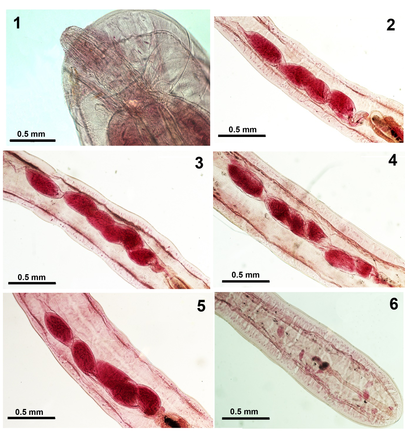
Figures 1-6: Echinorhynchus lateralis from Salvelinus fontinalis in Canada. 1. Proboscis of a female specimen. 2-5. Variations in the organization of cement glands of 5 selected specimens. 6. Detail of the female reproductive system.
Measurements and counts of our specimens are detailed in Table 2 compared to those of Leidy [8], Richardson [4], Sandeman and Pippy [7], Golvan [5], and Arai [6]. Golvan’s [5] account was a rendition of that of Leidy [8] and that of Arai [6] a blending of those of other authors combined. Sanderman, et al. [7] specimens from Newfoundland were considerably larger than all others on practically all counts except the egg size (Table 2). Our specimens provided more descriptive information as follows. The size of lemnisci exceeded that reported by other observers, sexual differentiation was demonstrated for the first time in the size of proboscis, hooks, and receptacle, and the size of each testis was provided separately instead of being combined. Some of the variations in the organization number of cement glands (Figs. 2-5) and the female reproductive system (Fig. 6) are shown.
Specimens: Harold W. Manter Lab., University of Nebraska State Museum, Lincoln HWML coll. no. 217509.
| Source | Leidy (1951), Richardson (1936), Golvan (1969) | Sanderman & Pippy (1967) | Arai (1989) based on other accounts | This paper |
|---|---|---|---|---|
| Geography | Lake Edward, Quebec, New York | Newfoundland | Quebec, Ontario, Newfoundland, Labrador, New Brunswick | Canada |
| Host | Salvelinus fontinalis (Mitchell) | Salvelinus fontinalis (Mitchell) & 6 salmonid & coregonid species | Salvelinus fontinalis (Mitchell) & 17 other species | Salvelinus fontinalis (Mitchell) |
| Sample size | Unknown | 41 males, 47 females | Unknown | 6 males, 7 females |
| Figures | 8-11 & 171-172 | 11 | 14 a-c | 1-6 |
| Characters | ||||
| Trunk L X W (mm) male | 6.0-7.0 X 0.7-0.9 | 3.0-17.5 X 0.40-1.6 | 2.0-17.5 X 0.4-1.6 | 6.9-8.7 X 0.7-0.9 |
| Trunk L X W (mm) female | 10.0-14.0 X 1.0 | 4.5-31.0 X 0.3-2.0 | 10.0-14.0 X 1.0 | 6.2-15.7 X 0.7-1.1 |
| Proboscis L X W (male) | 750 X --- | 435-813 X --- | 700-750 X --- | 614-728 X 177-208 |
| Proboscis L X W (female) | 750 X --- | 435-813 X --- | 700-750 X --- | 676-936 X 177-230 |
| Hook rows X H/row | 11-12 X 6 circular | 12-17 X 10-14 (females), 12-18 X 10-12 (males) | 12-18 X 10-12 | --- X up to 12-13 |
| Ant. hook L | 32-40 | 32-65 (females), 25-57 (males) | 25-27 | --- |
| Middle hook L | 36-48 | 35-63 (females), 25-58 (males) | 25-58 | 52-58 (females), 47-62 (males) |
| Post. hook L | 28 | 28 (both sexes) | 23-55 | 32-42 (females), 26-36 (males) |
| Hook roots | --- | As long as blades | As long as blades | --- |
| Recept. (female) (mm) | 0.83-1.25 X 0.21-0.25 | |||
| Recept. (male) (mm) | --- | 0.46-1.38 X --- | 0.46-1.38 X --- | 0.77-1.05 X 0.17-0.25 |
| Lemnisci L vs. recept. L L X W (male) L X W (female) | 1/2 to 3/4 | Ca. 1/2 | 1/2 to 3/4 | 1/2 to 125% 625-950 X 175-225 625 X 1,200 X 200-250 |
| Ant. testis L X W (mm) Post. testis L X W (mm) | 0.7-1.0 X 0.3-0.4 0.7-1.0 X 0.3-0.4 | 0.29-1.68 X 0.12-0.97 0.29-1.68 X 0.12-0.97 | 0.29-1.68 X 0.12-0.97 0.29-1.68 X 0.12-0.97 | 0.47-1.07 X 0.27-0.37 0.52-0.95 X 0.25-0.42 |
| Cement glands | 6 (1+2,3+4+5,6) | Variable | 6 (variable) | 4-6 contiguous or variable |
| Bursa L X W | --- | --- | --- | 500-625 X 350-425 |
| Female rep. syst. (mm) | --- | --- | --- | 1.04-1.35 |
| Eggs L X W | 88-112 X 20, polar prolongation | 65-110 X ---, polar prolongation | 65-112 X 20, polar prolongation | 78-83 X 16-21, polar prolongation |
Table 2: Comparative morphometrics of various Canadian populations of Echinorhynchus lateralis.
Echinorhynchus salmonis Müller, 1784 (=Echinorhynchus coregoni Linkins in Van Cleave, 1919; MetEchinorhynchus salmonis (Müller, 1784) Petrochenko, 1956 (Echinorhynchidae) (Figs. 7-11)
We obtained 16 specimens of E. salmonis from the posterior intestines of 3 of 14 (6 males & 8 females) lake herring (cisco) Coregonus artedii Lesueur (Salmonidae) examined in Lake Superior in June 1981. Two specimens were long large females and the other 14 were markedly smaller adults all distended medially as is characteristic of worms described from salmonid hosts, e.g., bloater, Coregonus hoyi (Gill) compared to the slender forms observed in non- salmonids, e.g., rainbow smelt Osmerus mordax (Mitchill) (Osmeridae) in Lake Michigan by Amin and Redlin [9]. Coho salmon Oncorhynchus kisutch Walbaum is the major salmonid fish species in Lake Superior which is also known to be heavily infected with E. salmonis in Lake Michigan (prevalence 100%; max. 796/fish) (Amin and Burrows, p 327) [10].
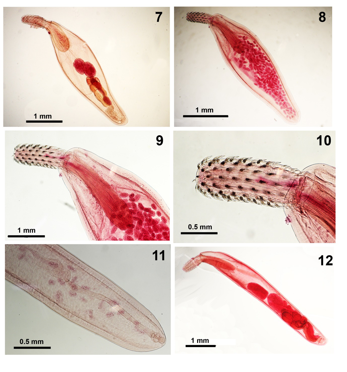
Figures 7-11: Echinorhynchus salmonis from Coregonus artedii in Lake Superior. 7. A whole mount of a young mature male specimen. 8. A female specimen in the ovarian ball stage. 9. A higher magnification of the female in Fig. 8 showing the proportional sizes of the proboscis vs. the receptacle. 10. A higher magnification of the proboscis of the same female specimen showing the distribution of hooks. 11. The reproductive system of another female specimen. Figure 12. A whole mount of a male specimen of Acanthocephalus lucii from Perca fluviatilis in the Ubort River, Ukraine. Note the differential sizes and distribution of the various organs.
Echinorhynchus salmonis is known in large bodies of water in the former USSR, Europe, and North America in salmonid and other fish species and has been described by many authors. Meyer [11] and Golvan [5] described it from Lake Onega, USSR using 3 figures of a male, a proboscis, and an egg after Lühe [12]. Petrochenko [13] listed 39 fish host species, described specimens based on his material also from Lake Onega, among other locations, and used the same 3 figures of Lühe [12]. Arai’s [6] description was a modification of those created by other authors including Van Cleave [14], Petrochenko [13], Yamaguti [15], and Golvan [5]. The most updated and comprehensive description of E. salmonis was provided by Amin, et al. [9] who provided regression analysis of anatomical variability as affected by worm sex, growth, and development, and host species and accounted for their taxonomic ramifications considering the full range of variation in 24 characters in 403 and 315 worms from C. hoyi and O. mordax in Lake Michigan.
Our two large females from lake herring, C. artedii in Lake Superior were collected from an 18 cm long male herring individual on June 7, 1981. The 14 small specimens were collected from a 12 cm long male and 35 cm long female herring in June and January 1981. Measurements of two large females (and 5 smaller females) are: trunk 12.50-15.00 X 1.37-1.57 mm (2.92-4.17 X 0.72-1.25 mm), proboscis 832-884 X 343-354 (700-900 X 275-301), hooks in 14-16 rows each with 9-10 hooks, receptacle 1.55-1.65 X 0.35-0.42 mm (1.12-1.30 X 0.30-0.40), lemnisci 1.25-1.45 X 0.25-0.30 mm (0.87-1.07 X 0.15 mm), reproductive system 1.62-2.02 mm (1.87 mm), eggs fusiform 73-78 X 16-21with polar prolongation of middle membrane. Measurements of 6 small males are: trunk 2.37-3.40 X 0.62-0.85 mm (Fig. 7), proboscis 600-697 X 229-261, hooks in 14 rows each with 11 hooks, receptacle 0.82-1.17 X 0.27-0.32 mm, lemnisci 0.67- 1.00 X 0.17-0.22 mm, Anterior testis 325-625 X 275-375, posterior testis 350-525 X 250-425, 6 cement glands 225- 280 X 156-200, 2 cement gland ducts surround bulb-shaped Saefftigen’s pouch. The small females’ measurements were: trunk 2.92-4.14 X 0.72-1.25 mm (Fig. 8), proboscis 700-900 X 275-301 (Figs. 8-10), receptacle 1.12-1.30 X 0.30-0.40 mm, lemnisci 0.87-1.07 X 0.15-0.22 mm, reproductive system 1.87 mm long (Fig. 11), and eggs 73-78 X 16-21.
Specimens: Harold W. Manter Lab., University of Nebraska State Museum, Lincoln HWML coll. no. 217510. Acanthocephalus lucii (Müller, 1776) Lühe, 1911 (= Echinorhynchus angustus Rudolphi, 1802) (Paracanthocephalidae) (Figs. 12-16) We have collected 5 males and 5 females of A. lucii from the European perch Perca fluviatilis Linn. in Ubort River, Ukraine, from where it was also collected by Rubtsova, et al. [16]. Acanthocephalus lucii is one of the most widely distributed species of acanthocephalans infecting freshwater fishes of Europe. Since its original description in Lühe [12], it has been described by many observers including Meyer [11], Lundström [17], Petrochenko [13], Yamaguti [15], Golvan [5], and Olburs [18], among others, most of whom used the brief but adequate description and line drawings of Müller [19] in Lühe [12]. Redescriptions were provided by Kostylew [20], Meyer [11], and Markowski [21]. Amin, et al. [22] provided the first SEM description of over 100 specimens of this species that were collected from the same host, P. fluviatilis in England, Finland, and Germany. Our 10 specimens from Ukraine fit well within the parameters originally described by Müller [19] in Lühe [12] which included measurements of the trunk, neck, proboscis receptacle, and eggs, proboscis hook formula, the shape of hook roots, lemnisci and the male reproductive system and also included good figures of a male, a female, a proboscis, rooted proboscis hooks, female reproductive system, and an egg. Our 10 Ukrainian specimens were compatible with the 12 SEM images of our specimens from England, Finland, and Germany [22] that included the proboscis, hooks, neck sensory pit, micropores, reproductive orifices, eggs, and bursa. In this presentation we offer a new light microscopy perspective of A. lucii showing additional features from 5 males (3.17-5.62 mm X 0.45-0.75 mm) (Figs. 12-15) and 5 females (11.12-18.32 mm X 0.87-1.10 mm) (Fig. 16) from Ukraine.
Specimens: Harold W. Manter Lab., University of Nebraska State Museum, Lincoln HWML coll. no. 217511.
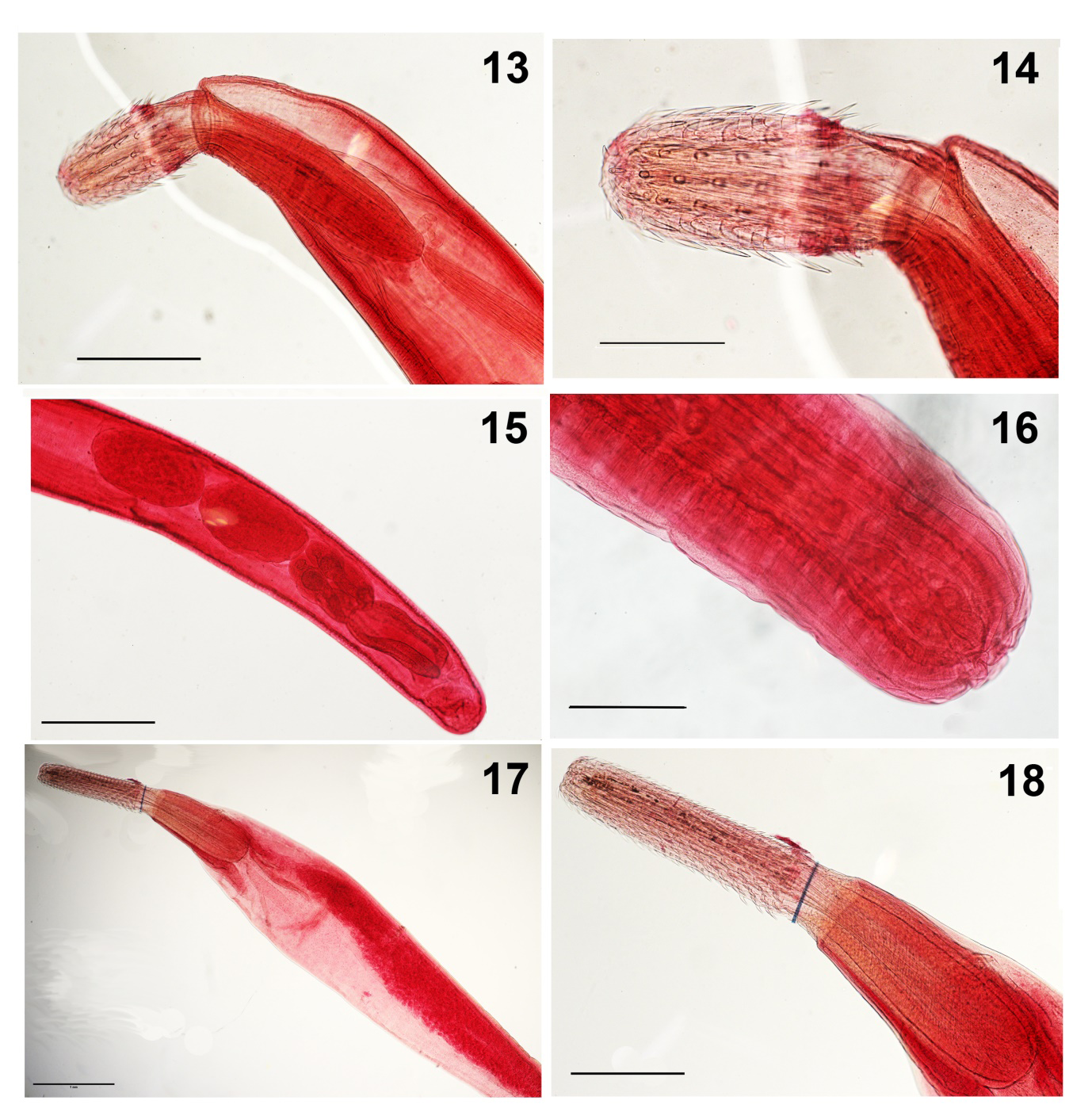
Figures 13-16: Acanthocephalus lucii from Perca fluviatilis in the Ubort River, Ukraine. 13. Anterior portion of a specimen showing the shape and proportional sizes of the proboscis vs. the receptacle. 14. A higher magnification of the proboscis of specimen in Fig. 13. 15. Detail of the male reproductive system. Note the two cement gland ducts. 16. The posterior part of the female reproductive system showing the terminal gonopore and the inflated vagina Figures 17 & 18. Acanthocephalus rhinensis from Anguilla anguilla in Lake Piediluco, Italy showing a whole female and the anterior portion of the same worm emphasizing the proboscis and the receptacle.
Acanthocephalus rhinensis Amin, Thielen, Mïnderie, Taraschewski, Sures, 2008 (Paracanthocephalidae) (Figs. 17-20)
Acanthocephalus rhinensis is known from only two populations collected from the same host species, the European eel A. anguilla. A small population yielding 9 specimens (4 males, 5 females) collected from 3 of 390 eels examined for parasites between 1995 and 2005 in the main Rhine River near the city of Karlsruhe, Germany was used for the description by Amin, et al. [23]. The males were 7.25-8.55 mm long by 1.00-1.07 mm wide and the females were 9.12-11.70 mm long by 1.25-1.55 mm wide. The larger population yielded 1,076 adults collected from 32 infected of 37 examined eels in Lake Piedliuco in Central Italy that appeared to “correspond in size and dimensions” to those in the original description according to Dezfuli, et al. [24]. The two localities are not geographically connected leaving open the possibility of introductions. A small sample of 5 males and 3 females of the Italian specimens was provided to Amin for confirmation of identification that, however, included relatively smaller specimens: males 2.82-4.50 mm long by 0.62-0.87 mm wide (Fig. 20) and females 5.75-7.75 mm long by 0.65-1.05 mm wide (Fig. 17). The long cylindrical proboscis and receptacle are featured in Figs. 18 & 19.
Dezfuli, et al. [24] were preoccupied by the the absence in their Italian specimens of the red brown belt at the anterior trunk of our German specimens (Fig. 8) and did not mention the key to European species of Acanthocephalus Koelreuther, 1771 provided by Amin, et al. [23] which distinguished A. rhinensis from all other species of Acanthocephalus in Europe based on proboscis armature, shape and size of testes, and eggs.
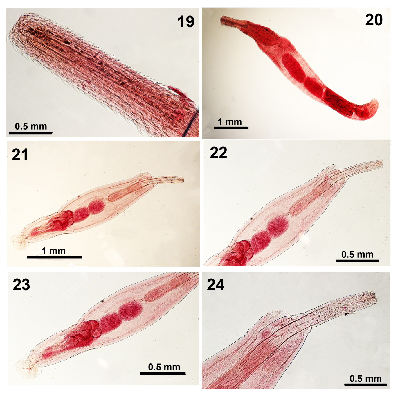
Figures 19 & 20: Acanthocephalus rhinensis from Anguilla Anguilla in Lake Piediluco, Italy shows a higher magnification of the proboscis and the male reproductive system. Figures 21-24. Acanthocephalus tahlequahensis from Etheostoma punctulatum in Illinois River tributaries, Oklahoma. 21. A paratype male. 22, 23. The enlarged anterior and posterior portions of the paratype male in Fig. 21. 24. A higher magnification of the anterior portion of the same male specimen shows the smaller size of the lemnisci compared to the receptacle.
The preoccupation of Dezfuli, et al. [24] with the absence of the brown-red belt failed to recognize the known fact that coloration in the Acanthocephala is often related to host diet usually involving carotenoids that have been identified by Barret and Butterworth [25] as esterified astaxanthin in Polymorphus minutus Goeze, 1782. Other color patterns have been associated with Pomphorhynchus bulbocolli Linkins in Van Cleave, 1919 infecting white suckers Catostomus commersoni (Lacépède) (see Amedjo and Holmes [26] [26] and Awachie [27]) recognized 4 color morphs of Echinorhynchus truttae Schrank, 1788 in Salmo trutta Linn. The diet of eels in the Rhine may well be different than in disjunct Italian lakes.
Amin, et al. [23] provided 7 informative line drawings of a male, a female, a proboscis, a female reproductive system, hooks and roots, and an egg. Dezfuli [24] provided SEM images of a proboscis and hooks and discussed the cycle of infection of cystacanths in the amphipod intermediate host Echinogammarus tibaldii (Pinkster & Stock, 1970). In this presentation, we provide additional perspectives of A. rhinensis as seen by light microscopy, for the first time (Figs. 17-20).
Specimens: Harold W. Manter Lab., University of Nebraska State Museum, Lincoln HWML coll. no. 217512.
Acanthocephalus tahlequahensis Oetinger and Buckner, 1976 (Paracanthocephalidae) (Figs. 21-25)
Acanthocephalus tahlequahensis was described from 4 species of fish in the Fox Creek, an intermittent tributary of the Illinois River in Cherokee County, Oklahoma. We acquired 4 specimens (2 males, 2 females) from the type host, the stippled darter Etheostoma punctulatum (Agassiz). The strippled darter is a geographically restricted species and the only one from which gravid females were originally found. Stratified histograms demonstrated that A. tahlequahensis, along with Acanthocephalus alabamensis Amin and Williams, 1983 fall into a natural group distinct from that of the more widely distributed species Acanthocephalus dirus (Van Cleave, 1931) [14] in North America (Amin and Huffman) [28]. From an evolutionary standpoint, these two southern species exhibited “new restricted distributions (in Oklahoma and Alabama)” or “exhibiting relictual distribution” [29].
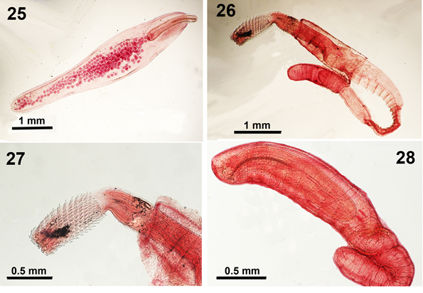
Figures 25: A whole female in the ovarian ball stage of Acanthocephalus tahlequahensis from Etheostoma punctulatum in Illinois River tributaries, Oklahoma. Figures 26-28. Cystacanths of Serrasentis sagittifer from Pterois volitans in the Philippines.
Acanthocephalus tahlequahensi appears to be somewhat more widespread in the Illinois River. McAllister, et al. [30, 31] also found it in Etheostoma radiosum Hubbs & Black, among other fish species from other tributaries of the Illinois River in adjacent Arkansas. Reports of its presence in various fish species and in the isopod Caecidotea communis (Say) in the coastal New Jersey Pinelands watershed in the context of ecosystem interactions and food webs by Hernandez and Sukhdeo [32] Sukhdeo [33], and Paseka [34] did not make any reference to the species description, substantiate its morphological identity, or show any connection to the Illinois River.
Our A. tahlequahensi males and females are paratypes gifted by David Oetinger that measured 2.25-2.75 X 0.55- 0.68 mm (Figs. 21-24) and 3.60-4.10 X 0.65-0.82 mm (Fig. 25), respectively. All other measurements fit within the range provided in the original description. Oetinger & Buckner [35] included 5 informative line drawings of a whole male, 2 proboscides, a female reproductive system, and an egg. Our specimens were sufficiently informative that our light microscope images provided additional perspectives not seen in the original line drawings such as a whole female and proboscis hook roots, or described in the original text.
Specimens: Harold W. Manter Lab., University of Nebraska State Museum, Lincoln HWML coll. no. 217513.
Serrasentis sagittifer (Linton, 1889) Van Cleave, 1923 juvenile (Rhadinorhynchidae) (Figs. 26-26)
The names Serrasentis Van Cleave, 1923, and Serrasentis sagittifer (Linton, 1889) Van Cleave, 1923 had undergone considerable nomenclature changes over the years and survived a few redescriptions under different names. Paul Sikkel kindly provided us with 10 small encysted juveniles, that appeared to be identifiable as S. sagittifer, from the body cavity of red lionfish Pterois volitans (Linn.), a paratenic host, in the Philippines in 2011. These are new host and locality records. The two most recent and significant publications on S. sagitiffer are those of Barton, et al. [36] and Amin & Heckmann [37]. Barton, et al. [36] redescribed the species morphologically using 11-line drawings that depicted errors in the presentation of posterior hooks and roots and male and female gonopores and used 6 SEM images. Barton, et al. [36] also provided baseline 18S rDNA, 28S rDNA, and cox1 sequence data and qualified the synonymies and host specificities as related to host factors (Barton et al., 2018). Amin & Heckmann [37], on the other hand, detailed the history of the concept S. sagittifer and its synonyms, described juveniles and adults and their geographical distribution that was shown to be correlated with the distribution of its major adult host Rachycentron canadum (Linn.) in Atlantic North and South America, Atlantic West Coast of Africa, The Indian Ocean to East Africa, the Red Sea and the Arabian Gulf, The Indo-Pacific Ocean in Australia, Indonesian islands, South China Sea and Vietnam, and unusual localities in California, Oregon, and Washington. Descriptions from these regions were compared and evaluated for variabilities. Additionally, Amin and Heckmann [37] provided 17 SEMs of the morphology of this species for the first time and new accounts of the micropores and Energy Dispersive X-ray analysis (EDXA) showing 3 spectra.
In this presentation, we offer a few light microscope images of encysted juveniles of S. sagitiffer (Figs. 26-28) from the newly reported paratenic host P. volitans from the Philippines for the first time.
Specimens: Harold W. Manter Lab., University of Nebraska State Museum, Lincoln HWML coll. no. 217514.
Acknowledgments
This project was partially supported by an ongoing institutional grant from the Parasitology Center, Inc., Scottsdale, Arizona, USA. We are immensely grateful to the many collaborators who contributed the study materials that made the execution of this work possible. We are thankful to Drs. Yuliya Kutsokon and Mykola Shcherbatiuk, National Academy of Science of Ukraine for their cooperation with the fish collections. Collaborators simply shared research material as part of joint research understandings. We also gratefully recognize the help of Dr. Gabor Racz., Collection Manager, Harold W. Manter Laboratory of Parasitology, University of Nebraska-Lincoln for kindly accessing and cataloging our specimens.
References
-
Amin OM, Rubtsova NYu (2023a) New Annotated Records of Helminth Parasites, Mostly from North America, with Light Microscopy. I. Trematoda (Digenea), Cestoda, Nematoda. Int J Zoo Animal Biol 6(5): 000507.
-
Amin OM, Rubtsova NYu (2023b) New Annotated Records of Helminth Parasites with Light Microscopy II. Acanthocephala (Polymorphida). Int J Zoo Animal Biol 6(6): 000530.
-
Amin OM, Rubtsova NYu (2023c) New Annotated Records of Helminth Parasites with Light Microscopy. III. Acanthocephala (Archiacanthocephala and Eoacanthocephala). Int J Zoo Animal Biol 2023, 6(5): 000507.
-
Richardson LR (1936) Observations on the parasites of speckled trout in Lake Edward, Quebec. Trans Amer Fisher Soc 66: 343-356.
-
Golvan Y (1969) Systématique des Acanthocephales (Acanthocéphala Rudolphi, 1801), L’ordre des Palaeacanthocephala Meyer, 1931, La superfamille des Echinorhynchidea (Cobbold, 1876) Golvan et Houin 1973. Mémoires du Muséum Natl d’histoire Nat 47: 1-373.
-
Arai HP (1989) Acanthocephala, p 1-90. In: Margolis L, Kabata Z (Eds.), Guide to the Parasites of Fishes of Canada. Part III. Can Spec Publ Fish Aqua Sci 107: 95.
-
Sandeman IM, Pippy JHC (1967) Parasites of freshwater fishes (Salmonidae and Coregonidae) of insular Newfoundland. J Fisher Res Bd Canada 24 (9): 1913- 1943.
-
Leidy J (1851) Contributions to Helminthology. Proc Acad Nat Sci Philadelphia 5: 205-209.
-
Amin OM, Redlin MJ (1980) The effect of host species on growth and variability in _Echinorhynchus salmonis_ Müller, 1784 (Acanthocephala: Echinorhynchidae), with special reference to the status of the genus. Syst Parasitol 2: 9-20.
-
Amin OM, Burrows JM (1977) Host and seasonal associations of _Echinorhynchus_ _salmonis_ (Acanthocephala: Echinorhynchidae) in Lake Michigan fishes. J Fish Res Board Can 34: 325-331.
-
Meyer A (1932) Acanthocephala. In: Bronn HG (Ed.), Klassen und Ordnungen des Tier-Reichs, Akad Verlag MBH, Leipzig 4: 1-332.
-
Lühe M (1911) Acanthocephalen. Die Slsswasserfauna Deutschlands, Heft l6, Jena.
-
Petrochenko VI (1956) Acanthocephala of Domestic and Wild Animals. Acad Sci USSR pp: 465.
-
Van Cleave HJ (1924) A critical study of the Acanthocephala described and identified by Joseph Leidy, Proceedings of the Academy of Natural Sciences, Philadelphia 76: 279-334.
-
Yamaguti S (1963) Acanthocephala. In Systema Helminthum. Wiley Interscience, New York, USA. 5: 1-423.
-
Rubtsova NYu, Kutsokon YuK (2018) First note on fish parasites in Polissky Nature Reserve, Northern Ukraine. Vestnik Zoologii 52(1): 53-58.
-
Lundström A (1942) Die Acanthocephalen Schwedens. Mit Ausnahme der Fischacanthocephalen von Süsswasserstandorten. Doctor’s thesis, Sci Univ Lund, Sweden, pp: 238.
-
Olburs C (1978) Gastrointestinal helminths in pike, _Esox_ _lucius_ L., from fresh and brackish water. Inform. från Sötvattens-laboratoriet Drottningholm No 13, Sweden, pp: 100.
-
Müller OF (1776) Zoologiae danicae prodromus sue animaleum daniaeet norvegiae indigenarum characters, nomina, et synonyma imprimis popularium pp: 282.
-
Kostylew NN (1916) Contributions to the acanthocephalan fauna of Russia. Ann Zool Mus Imper Acad Sci Petrograd 20: 389-394.
-
Markowski S (1933) Die Eingeweidewürmer der Fische des Polnischen Balticums (Trematoda, Cestoda, Nematoda, Acanthocephala). Arch d’Hydrobiol et d’Ichthyol 7: 1-58.
-
Amin OM, Heckmann RA, El Naggar AM (2011) Revisiting the morphology of _Acanthocephalus lucii_ (Acanthocephala: Echinorhynchidae) in Europe, using SEM. Sci Parasitol 12 (4): 185-189.
-
Amin OM, Thielen F, Münderle M, Taraschewski H, Sures B (2008) Description of a new echinorhynchid species (Acanthocephala) from the European eel, _Anguilla anguilla_, in Germany, with a key to species of Acanthocephalus in Europe. J Parasitol 94(6): 1299- 1304.
-
Dezfuli BS, Lui A, Squerzanti S, Lorenzoni M, Shinn AP (2012) Confirmation of the hosts involved in the life cycle of an acanthocephalan parasite of _Anguilla anguilla_ (L.) from Lake Piediluco and its effect on the reproductive potential of its amphipod intermediate host. Parasitol Res 110: 2137-2143.
-
Barrett J, Butterworth PE (1968) The carotenoids of _Polymorphus minutus_ (Acanthocephala) and its intermediate host, _Gammarus pulex_. Comparative Biochemistry and Physiology 27(2): 578-581.
-
Amedjo SD, Holmes JC (1989) Color, size, and maturation of _Pomphorhynchus bulbocolli_ (Acanthocephala) in white suckers, _Catopstomus commersoni_. J Parasitol 75 (5): 798-800.
-
Awachie JBE (1966) The development and life history of _Echinorhynchus truttae_ Schrank, 1788 (Acanthocephala). Journal of Helminthology 40: 11-32.
-
Amin OM, Huffman DG (1984) Interspecific variability in the genus _Acanthocephalus_ (Acanthocephala: Echinorhynchidae) from North American freshwater fishes, with a key to species. Proc Helminthol Soc Wash 51: 238-240.
-
Amin OM (1985) Hosts and geographical distribution of _Acanthocephalus_ (Acanthocephala: Echinorhynchidae) from North American freshwater fishes, with a discussion of species relationships. Proc Helminthol Soc Wash 52(2): 210-220.
-
McAllister CT, Connior MB, Font WF, Robison HW (2014) Helminth parasites of the Banded Sculpin, _Cottus_ _carolinae_ (Scorpaeniformes: Cottidae), from Northern Arkansas, USA Comparative Parasitology 81: 203-209.
-
McAllister CT, Richardson DJ, Barger MA, Fayton TJ, Robison HW (2016) Acanthocephala of Arkansas, including new host and geographic distribution records from fishes. J Arkans Acad Sci 70: 26.
-
Hernandez AD, Sukhdeo MVK (2008) Parasites alter the topology of a stream food web across seasons. Oecologia 156: 613-624.
-
Sukhdeo MVK (2012) Where are the parasites in food webs?. Parasites and Vectors 5(1-17): 239.
-
Paseka RE (2018) Linking host-parasite interactions and ecosystem processes with energy and elements. Ph D dissert, Rutgers Univ, New Brunswick, New Jersey, USA, pp: 162.
-
Oetinger DF, Buckner RL (1976) _Acanthocephalus_ _tahlequahensis_ sp. n. (Acanthocephala: Echinorhynchidae) from the stippled darter, _Etheostoma_ _punctulatum_ (Agassiz), in northeastern Oklahoma. J Parasitol 62(2): 237-241.
-
Barton DP, Smales L, Morgan JAT (2018) A redescription of _Serrasentis_ _sagittifer_ (Rhadinorhynchidae: Serrasentinae) from _Rachycentron_ _canadum_ (Rachycentridae) with comments on its biology and its relationship to other species of _Serrasentis_. J Parasitol 104(2): 117-132.
-
Amin OM, Heckmann RA (2021) New perspectives on the distribution, morphology, and hook chemistry of _Serrasentis sagittifer_ (Linton, 1889) Linton, 1932 using SEM and Energy Dispersive X-ray analysis. Sci Parasitol 22(2-3): 88-111.
- California Red-Legged Frog and Non-Listed Amphibians Response to Non-Native Fish Removal
- Industrial Standardization of the Bio-OS: Algorithmic Codification of Resilience Engineering Guidelines and Version V8 Architecture
- Climate Variability and the Sustainability of Snail Farming in Nigeria: Past Trends, Present Challenges and Potential Outlook
- The Evaluation of the Surveillance System of Anthrax in Gilgit-Baltistan, Pakistan, 2018
- Natural Decline to Extinction of A New Zealand Rabbit Population
- Mitochondrial Bio-Logistics: Steering Co-Enzyme Q10 and Lycopene Synergies within the Science 4.0 Bio-OS Framework