Medial Epicondyle Fractures of Humerus in Children at the University Teaching Hospital of Yopougon
The purpose of this study is to evaluate the results of surgical treatment of medial epicondyle fractures in children. Patients and Methods: A retrospective study of 17 children was performed in the Pediatric Surgery Department at the Yopougon Hospital and University Center between January 2004 and December 2014. Children aged 3 to 15 years treated surgically for a fracture of medial epicondyle and had regular postoperative follow-up of more than three months were included. The average age was 11 years old. Fractures were divided according to the Marion and Faysse classification: stage II (n = 5), stage III (n = 3), stage IV (n = 9). We evaluated postoperative complications and sequelae. The evaluation of the functional results was based on the criteria of Hardacre. Results: The functional results were studied with a mean follow-up of 8 months. We found 70% good and very good results, 18% average results and 12% poor results. Postoperative complications were observed n = 3 (17.6%) of the cases. It was an operative wound infection n = 2 (11.7%), iatrogenic nerve damage n = 1 (5.9%). Sequelae were represented n = 3 (17.6%). This was an n = 2 elbow mobility deficit (11.7%) and the epitrochlear n = 1 protrusion (5.9%). Conclusion: Medial epicondyle fracture outcomes are associated with a relatively low rate of complications. It is important to stress the importance of prolonged surveillance because of the functional and morphological sequelae that they may cause.
Introduction
Fractures of the lower extremity of the humerus represent a pathology very frequently encountered in pediatric traumatology. Fractures of the medial epicondyle represent 10% of elbow fractures [1]. Its imperfect reduction may be responsible for aesthetic sequelae and disabling functional deficits [2]. The treatment is traditionally done urgently and obeys various orthopedic and / or surgical methods. However, their open treatment remains the only therapeutic method for medial epicondyle fractures displaced in our working conditions. This study aims to evaluate the results of surgical treatment of these fractures.
Patients and Methods
This is a retrospective study of the 23 children treated in the Pediatric Surgery Department at the Yopougon Hospital and University for a fracture of the medial epicondyle, from 2004 to 2014, in 11 years. Patients aged between 3 and 15 years undergoing surgery and having regular post-operative follow-up of more than three months were included. Cases treated orthopedically were not included in our study. The study covered 17 files. There were 14 boys (82.4%) and 3 girls (17.6%), a sex ratio of 4.7. The average age was 11 years extreme (3 and 15 years). Fractures were divided according to the Marion and Faysse classification (Table 1) [3]: stage II (n = 5), stage III (n = 3), stage IV (n = 9).
Stage I: absent or minimal movement, below and sometimes forward. Stage II: significant displacement with fragment drawn downwards sometimes up to the line of the articular line humero-cubital. Stage III: incarceration of the epitrochlea in the elbow joint. Stage IV: Fractured fracture associated with elbow dislocation.
Table 1: Classification according to Marion and Faysse [3]. The therapeutic delay, defined as the time between admission to the emergency department and admission to the operating theater, was on average 2.42 extreme days (1 and 5 days). All children underwent general anesthesia in the supine position, the upper limb on a tablet. After placing a tourniquet at the root of the arm, a medial cutaneous approach was performed, and then the ulnar nerve was isolated and placed on a lake. After the reduction, two identical Kirschner wires of diameter 1.5 and 1.8 millimeters were mounted to the electric motor (Figures 1a and 1b). The stability of the assembly was checked. The pins were then cut, bent and buried subcutaneously. The closure was done in two planes on a suction redon drain with an intradermal over lock. After removal of the withers, a plastered posterior splint was placed, the elbow bent at 90 °. On the third day, the redon drain was removed and a brachial-antebrachio-palmar cast was made for a period of thirty to forty-five days. At the end of this period, the spines were removed as outpatients.
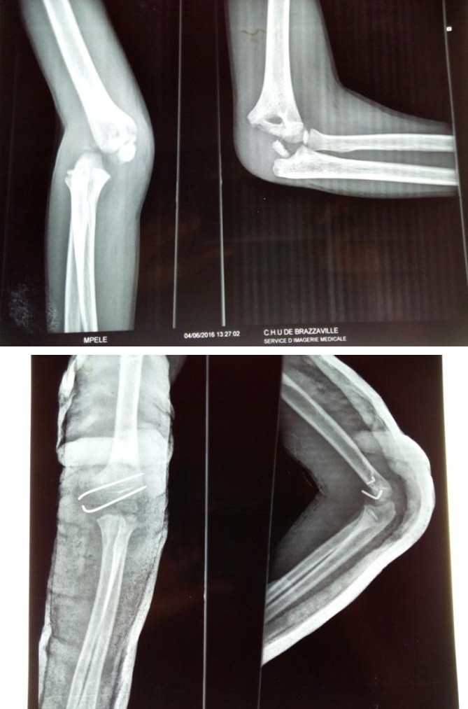
Figure 1a: Incarceration of the epitrochlea in the joint (stage 3).
Figure 1b: Post Operative Radiography. We looked for post-operative complications (iatrogenic nerve injury, infection, secondary displacement) and sequelae (protrusion of the epitrochlea, limitation of joint mobility, non-union, axis deviation). Our evaluation criteria were based on clinical and radiological data, represented by: - mobility of the elbow, based on the international score on the one hand (bending at 145°, extension at 0°, prono-supination at 90°) and evaluating the loss of amplitude compared to the healthy side of somewhere else. - The axial deflection and projection of the epitrochlée. - The unsightly surgical scar. - neurological disorders of the ulnar nerve.
- and finally, the radiological examination to evaluate the consolidation of the fracture site. These criteria meet the HARDACRE classification [2, 4] (Table 2). The results were satisfactory if they were very good, good or average.
| Mobility | Morphology | Complications |
| Stage 1(very good) | normal | Normal |
| Stage 2(good) | decrease(<10) | malalignment(<50) |
| Stage 3(way) | decrease(10-20) | malalignment(>50) |
| Stage 4(bad) | decrease(>50) | malalignment(>50),neurological signs,Pain,non-union |
Table 1: Classification according to Marion and Faysse [3].
Results
The overall results were satisfactory n = 15 (88%) cases evaluated after an average follow-up of 8 extreme months (3 and 36 months). According to Hardacre's evaluation criteria, we found n = 12 (70%) good and very good results, n = 3 (18%) average results and n = 2 (12%) poor results. Postoperative complications were observed n = 3 (17.6%). N = 2 (11.7%) cases of operative wound infection, n = 1 (5.9%) cases of iatrogenic nerve damage (ulnar nerve). Sequelae were represented n = 3 (17.6%). This was a limitation of the elbow mobility n = 2 (11.7%): the first case was observed in the patient with a stage II fracture with an extension deficit between 10-20° and the second case in a patient with a stage IV fracture with a flexion deficit of the elbow between 20 - 30 °. These 2 cases were treated by passive rehabilitation. The protrusion of the epitrochlea was noted n = 1 (5.9%) in a patient who had a stage III fracture. No cases of pseudarthrosis or axial deviation were observed. Discussion The limitations of our study were related to its retrospective nature and a low-rate sample. This study only involved large displacement fractures treated in a stereotyped manner using a single method. The medial epicondyle fracture treatment results are good and very good, they vary between 66.7 to 100% [2, 5, 6, 7, 8], thus joining the results obtained in our series where they represent 70%. The average and poor results reported by the authors vary between 20 and 28% of cases and 30% in our study [2, 5]. Our attitude was open-ended reduction in first intention. This treatment allowed us to obtain 88% of satisfactory results. Postoperative complications were dominated by infections. Superficial suppurations have been reported and treated with local care and adapted antibiotherapy that has evolved favorably as in our study [9, 10]. Bede, et al. observed one case of postoperative arthritis and one case of abscess [11]. Iatrogenic nerve damage (ulnar nerve) was observed in our study, having evolved favorably after 9 months, as pointed out by Lee, et al. [12]. This damage is due to traction on the nerve during reduction maneuvers. No cases of secondary displacement were noted. This could be explained by the good stability of the assembly. The limitation of the articular amplitudes of the postoperative elbow essentially concerns flexion and extension movements. A mobility deficit has been reported, as shown in our series [5, 7, 8, 12, 13, 14]. Dunn, et al. noted a correlation between mobility limitation and plaster duration, especially when it exceeds 5 weeks [15]. The projection of the epitrochlée has been observed by several works [2, 5], it is inconvenient from the aesthetic point of view. Pseudarthrosis secondary to fractures is rare. It is favored by insufficient reduction and secondary displacement [15]. The absence of pseudarthrosis in our series is explained by a good open surgical reduction and stability of the focus. Unlike in some studies, [8, 9] have been observed cases of nonunion. Axial deviation has been observed [3]. We did not note this complication; this could be explained by a slight decline in our series.
Conclusion
Fractures of the medial epicondyle are benign fractures whose prognosis depends on well-conducted treatment. Their results are accompanied by a relatively low rate of complications. It is important to stress the importance of prolonged surveillance because of the functional and morphological sequelae that they may cause.
References
-
Abuamara S, Lechevalier J (2002) Fractures of the medial epicondyle. In: JM Clavert, C Karger, Lascombes P, JN Ligier, JP Metaizeau (Eds.), Fractures of the child, Pediatric Orthopedics Study Group (GEOP). Montpellier Edition Sauramps Medical, pp: 125-130.
-
Yousri B, El Andaloussi M (1999) Fractures of the epitrochlea in children. The hand 4 (2): 71-77.
-
Marion J, Faysse R (1962) Fractures of the epitrochlea. Rev Chir Orthop 48: 447-470.
-
Fowles JV, Rizkallah R (1976) Intra-articular injuries of the elbow: pitfalls of diagnosis and treatment. Can Med Assoc J 114 (2): 125-131.
-
El Andaloussi Y, Yoursi B, Aboumaarouf M, El Andaloussi M (2006) Medial epicondyle fractures in children. Chir hand25 (6): 303-308.
-
Lawrence JT, Patel NM, Macknin J, Flynn JM, Cameron D, et al. (2013) Return to competitive sports after medial epicondyle fractures in adolescent athletes: results of operative and nonoperative treatment. Am J Sports Med 41(5): 1152-1157.
-
Bulut Erken Hy, Tan E, Ofluoglu, Yildiz (2005) Treatment of medial epicondyle fractures accompanying elbow dislocations in children. Acta Orthop Traumatol Turc 39(4): 334-340.
-
Rasool MN (2004) Dislocation of the elbow in children. J Bone Joint Surg 86(7): 1050-1058.
-
Ip D, Tsang WL (2007) Medial humeral epicondylar fracture in children and adolescents. J Orthop Surg (Hong Kong) 15(2): 170-173.
-
Hines RF, Herndon WA, Evans JP (1987) Operative treatment of medial epicondyle fractures in children. Clin Orthop Relat Res 223: 170-174.
-
Bede WB, Lefebvre AR, Rosman MA (1975) Fractures of the medial humeral epicondyle in children. Can J Surg 18(2): 137-142.
-
Lee HH, Shen HC, Chang JH, Lee CH, Wu SS (2005) Operative treatment of displaced medial epicondyle fractures in children and adolescents. J Shoulder Elbow Surg 14(2): 178-185.
-
Farsetti P, Potenza V, Caterini R, Ippolito E (2001) Long-term results of treatment of fractures of the medial humeral epicondyle in children. J Bone Joint Surg 83(9): 1299-1305.
-
Kamath AF, Baldwin K, Horneff J, Hosalkar HS (2009) Operative versus non-operative management of pediatric medial epicondyle fractures: a systematic review. J Child Orthop 3(5): 345-357.
-
Duun PS, Ravn P, Hansen LB, Buron B (1994) Osteosynthesis of medial humeral epicondyle fractures in children. 8 year follow-up of 33 cases. Acta orthop scand 65(4): 439-441.
- Return to Work Among Manual Workers After the Latarjet Procedure: A Cohort Study of 43 Patients
- Refractory Pelvic Collection Following Modified Stoppa Approach for Both-Column Acetabular Fracture Fixation: A Case Report
- Comparative Study of Dynamic Knee Phenotypes Under Loaded and Unloaded Conditions: Clinical Impact
- Locked Intramedullary Nailing of the Tibia Using a Humeral Nail: A Care Case Report
- Subtalar Dislocation: About a Case Report
- Surgical Site Infection in Orthopedics in a Country with LimitedResources: Indications, Treatment and Results