Factors Predicting Avascular Necrosis After Closed Reduction and Internal Fixation of Displaced Subcapital Fractures of the Hip
Background: The main complications following closed reduction and internal fixation of displaced intracapsular femur fractures (DICFF) are avascular necrosis (AVN) and nonunion. Little is known about the factors that predict these complications. The purpose of this study was to assess the clinical and radiological risk factors of failure following closed reduction and internal fixation of DICFF among non-elderly patients. Methods: We retrospectively reviewed patients under the age of 65 years who underwent closed reduction and internal fixation of DICFF in our center from January 2007 to January 2018. The extracted data included baseline characteristics and the fixation method. Postoperative radiographs were obtained during follow-up visits in order to evaluate complications, such as AVN and nonunion. Results: In total, 90 patients were included in the study, with a mean follow-up of 6.5 years. Garden III fractures were associated with significantly lower rates of AVN (odds ratio [OR] = 0.304, 95%confidence interval [CI]: 0.101-0.916) compared to Garden IV fractures. Neutral and varus reduction positions had significantly higher rates of AVN (OR = 7.182, 95%CI: 1.95126.446) and (OR= 5.560, 95%CI: 1.130-27.351), respectively, compared to the valgus position. Longer surgery procedures were associated with a higher risk of AVN (OR = 1.018, 95%CI: 1.006-1.031). Conclusions: Both varus and neutral reduction positions as well as Garden IV classification served as predictive factors for failure following closed reduction and internal fixation of DICFF. The type of fixation (CCS vs Targon plate) and the Pauwel angle had no significant effect on the rates of AVN or nonunion following fracture reduction.
Introduction
Hip fractures are a growing concern for healthcare systems worldwide and are responsible for a major decline in quality of life [1]. The incidence of femoral fractures is increasing, and it is estimated that there could be up to 4.5 million cases yearly by 2050 [2]. Anatomically closed reduction and internal fixation are mandatory in order to preserve the blood supply of the femoral head in patients younger than 65 years of age who sustained displaced intracapsular femur fractures (DICFF). The main complications following closed reduction and internal fixation of DICFF are avascular necrosis (AVN) and nonunion of the femoral head, with rates of 16% and 33%, respectively, having been reported in a meta-analysis [3].
While several studies have examined the complications that can arise from fixation and reduction, few studies have investigated the predictors for failure following this procedure. It has been shown that varus reduction, difficulty in achieving reduction, and fracture displacement were predictors for failure after surgery for DICFF [4–6]. The purpose of this study was to assess the clinical and radiological risk factors that determine failure following closed reduction and internal fixation of DICFF.
Methods
Our institutional research ethics board approved this retrospective study. A search of our institutional research database was performed to identify patients younger than 65 years of age who had undergone a closed reduction and internal fixation of DICFF between the January 2007 and January 2018. We excluded patients younger than 18 years, those who had a pathologic fracture, and those who were treated with primary total hip arthroplasty or hemiarthroplasty or did not meet the required two-year minimal follow-up period.
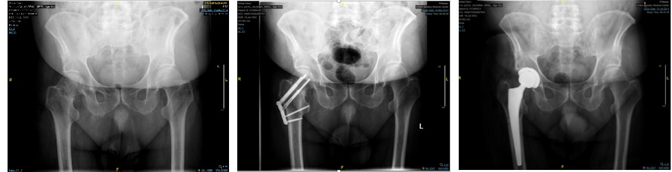
The data retrieved from the patients’ electronic medical records included baseline characteristics (e.g., sex, age), time from admission to surgery, surgery length, and fixation method. Preoperative radiographs that included an anteroposterior view of the pelvis and an anteroposterior of the hip were selected for evaluation in order to classify the fracture according to the Garden and Pauwel classifications [7, 8]. Garden Types I and II fractures are non-displaced, while Types III and IV fractures are displaced. Type III fractures represent complete fractures with partial displacement, indicated by a change in the angle of the trabeculae, and Type IV fractures represent complete fractures with complete displacement, leading to parallel orientation of the trabeculae [7]. The Pauwel classification categorizes fractures according to the angle of the fracture line and the horizontal axis, whereby fractures below 30 degrees are Degree I, those between 30-50 degrees are Degree II, and those of 50 degrees and above are Degree III [8].
All of the study patients were followed at routine postoperative visits at 6 weeks, 3 months, 6 months, 1 year, and yearly thereafter for a minimum of 2 years. Postoperative radiographs, including an anteroposterior view of the pelvis and anteroposterior and lateral views of the hip were routinely obtained during follow-up visits in order to evaluate union of the fracture as well as complications, such as nonunion and AVN. AVN was diagnosed when subchondral sclerosis or segmental collapse of the femoral head were revealed on simple radiographs. When AVN was clinically suspected but there were no corroborative signs on simple radiographs, the presence of AVN was determined by a magnetic resonance imaging (MRI) scan. Nonunion was defined by no progression in fracture healing after a period of 6 months [9].
Statistical Analysis
All of the statistical analyses were performed with IBM SPSS V24. We used mean and standard deviation (SD) or median and interquartile range (IQR) for numerical data and count (percentage) for categorical data. We applied logistic regression to assess the odds ratio (OR) for AVN and nonunion, and to account for the covariates of age at surgery, surgery length, sex, time to surgery, Garden and Pauwel classifications, reduction position, and type of fixation. Statistical significance was set at a p-value < 0.05.
Results
Between January 2007 and January 2018, 90 patients with DICFF were treated with closed reduction and internal fixation at our institution. There were 66 (73.3%) males. The mean age at surgery was 49 years (SD=8.92). Forty- one patients (45.6%) had a left-sided fracture (Table 1). The mean follow-up duration was 6.5 years (SD=2.4, range 2-11 years). Forty-nine hips were classified as Garden IV. The median time from emergency department admission to surgery was 7.5 hours (IQR = 5.2-9.95). Thirty-nine hips (43.3%) were fixated with three cannulated screws (CCS) and 51 hips (56.7%) were fixated with the Targon femoral neck (FN) Plate. The median surgery length was 71.5 (IQR=51-95) minutes, and the revision rate to total hip arthroplasty was 13.3% (n = 12).
| Sex | Men | 66 (73.3%) |
|---|---|---|
| Women | 24 (26.7%) | |
| Age at surgery (y) | Mean (SD) | 49 (8.92) |
| Garden n (%) | Garden III | 41 (45.5) |
| Garden IV | 49 (54.5) | |
| Time to surgery (H) | Median (IQR 25-75) | 7.5 (5.2-9.95) |
| Type of fixation n (%) | 3 Cannulated screws | 39 (43.3) |
| Targon FN plate | 51 (56.7) | |
| Avascular necrosis n (%) | 38 (42.22) | |
| Nonunion n (%) | 11 (12.22) | |
| Failure n (%) | 39 (43.33) | |
| Surgery length (m) | Median (IQR 25-75) | 71.5 (51-95) |
| Follow up time (y) | Mean (SD) | 6.5 (2.4) |
| Side n (%) | Left | 41 (45.6) |
| Right | 49 (54.4) | |
| Revision rate n(%) | 12 (13.3) |
Table 1: Results of 90 patients with DICFF in the year January 2007 and January 2018.
AVN of the femoral head
During the follow-up period, 38 patients (42.22%) were diagnosed with AVN of the femoral head (Table 2). The age at surgery (OR = 1.017, 95%CI 0.931-1.109), time to surgery (OR = 0.988, 95%CI 0.936-1.042), sex (OR = 0.86, 95%CI 0.264-2.811), type of fixation (OR = 0.744, 95%CI 0.186- 2.968), and Pauwel classification (p > 0.05) had no significant effect on the risk developing AVN after internal fixation.
Garden III fractures were associated with significantly lower rates of AVN compared to Garden IV fractures (OR= 0.304, 95%CI: 0.101-0.916). Neutral and varus reduction positions had significantly higher rates of AVN (OR= 7.182, 95% CI: 1.951-26.446 and OR= 5.560, 95% CI: 1.130-27.351, respectively), compared to valgus positions. Finally, longer surgery procedures were associated with a higher risk of AVN (OR= 1.018, 95%CI: 1.006-1.031).
- OR
- P-value
- 95% C.I.
- Lower
- Upper
- Sex
- 0.862
- 0.806
- 0.264
- 2.811
- Type of fixation Cannulated screws/Targon plate (Targon plate is reference)
- 0.744
- 0.675
- 0.186
- 2.968
- Garden 3 (garden 4 -reference)
- 0.304
- 0.034
- 0.101
- 0.916 reduction position - neutral (reduction position – valgus = reference)
- 7.182
- 0.003
- 1.951
- 26.446 reduction position - varus (reduction position – valgus = reference)
- 5.56
- 0.035
- 1.13
- 27.351 pauwel degree 2 (Pauwel degree 1 = reference)
- 0.437
- 0.512
- 0.037
- 5.165 pauwel degree3 (Pauwel degree 1 = reference)
- 0.836
- 0.887
- 0.07
- 9.921
- Age at surgery
- 1.017
- 0.714
- 0.931
- 1.109
- Time to surgery
- 0.988
- 0.65
- 0.936
- 1.042
- Surgery length
- 1.018
- 0.004
- 1.006
- 1.031
- 104.1), and time to surgery (OR=0.996, 95%CI: 0.984-1.006) had no significant effect on the OR of nonunion. However, age at surgery and surgery duration (OR=1.232, 95%CI: 1.030-
- OR
- P-value
- 95% C.I.
- Sex
- 1.971
- 0.499
- 0.275
- 14.13
- Type of fixation Cannulated screws/Targon plate
- 1.14
- 0.875
- 0.224
- 5.81 (Targon plate is reference)
- Garden 3 (garden 4 -reference)
- 0.558
- 0.463
- 0.118
- 2.65 reduction position - neutral (reduction position – valgus = reference)
- -
- -
- -
- reduction position - varus (reduction position – valgus = reference)
- 9.773
- 0.59
- 0.918
- 104.101 pauwel degree 2 (Pauwel degree 1 = reference)
- -
- -
- -
- pauwel degree3 (Pauwel degree 1 = reference)
- -
- -
- -
- -
- Age at surgery
- 1.232
- 0.22
- 1.03
- 1.473
- Time to surgery
- 0.996
- 0.415
- 0.987
- 1.006
- Surgery length
- 1.02
- 0.04
- 1.001
- 1.039
Table 2: OR of AVN.
Nonunion of the femoral head
During the follow-up period, 11 patients (12.22%) were diagnosed with nonunion of the femoral head (Table 3 and Figure 1). Sex (OR=1.971, 95%CI: 0.275-14.13), type of fixation (OR=1.140, 95%CI: 0.224-5.81), Garden III fractures (with Garden IV fractures as reference) (OR=0.558, 95%CI: 0.118-2.65), varus reduction (OR=9.773, 95%CI: 0.918-
Treatment failure of DICFF
Pauwel classification (p > 0.05), age at surgery (OR= 1.041, 95%CI: 0.978-1.108), and time to surgery (OR= 0.996, 95%CI: 0.988-1.005) had no significant effect on the OR of failure of surgical reduction and fixation. The duration of surgery (OR= 1.019, 95%CI: 1.006-1.032) and varus reduction (OR= 4.463, 95%CI: 1.107-17.997) or neutral positions (OR= 7.061, 95%CI: 1.917-26.014) had a significantly increased OR of failure.
During the follow-up period, 39 cases (43.33%) underwent failure of surgical reduction and fixation of DICFF and developed AVN or nonunion or both (Table 4). Sex (OR= 1.054, 95%CI: 0.327-3.392), type of fixation (OR = 1.502, 95%CI: 0.502-4.497), Garden III fractures (with Garden IV fractures as reference) (OR=0.394, 95%CI: 0.138-1.121), OR P-value 95% C.I.
Sex 1.054 0.93 0.327 3.392
Type of fixation Cannulated screws/Targon plate 1.502 0.467 0.502 4.497
(Targon plate is reference) reduction position - neutral (reduction position – valgus = reference) 7.061 0.003 1.917 26.014 Garden 3 (garden 4 -reference) 0.394 0.081 0.138 1.121
- reduction position - varus (reduction position – valgus = reference)
- 4.463
- 0.036
- 1.107
- 17.997 pauwel degree 2 (Pauwel degree 1 = reference)
- 0.503
- 0.5
- 0.68
- 3.704 pauwel degree3 (Pauwel degree 1 = reference)
- 1.213
- 0.85
- 0.164
- 8.969
- Age at surgery
- 1.041
- 0.206
- 0.978
- 1.108
- Time to surgery
- 0.996
- 0.411
- 0.988
- 1.005
- Surgery length
- 1.019
- 0.005
- 1.006
- 1.032
Table 3: OR of failure (AVN and nonunion combined).
Discussion
The purpose of this study was to assess the clinical and radiological risk factors that determine the failure of closed reduction and internal fixation for displaced intracapsular fractures of the hip in a population of patients under 65 years of age. The main reasons for treatment failure in DICFF were nonunion and AVN. The overall failure rate for the entire cohort of this study was 43.33%. The overall AVN rate for the entire cohort was 42.22%, and the overall nonunion rate was 12.22%. In addition, the overall conversion to total hip arthroplasty for the entire cohort at final follow-up was 13%.
A meta-analysis of 106 reports published by Lu-Yao, et al. [3] revealed nonunion and AVN rates of 33% and 16%, respectively, with up to 36% of patients undergoing revision surgery within 2 years from primary fixation surgery. Similarly, a meta-analysis published by Slobogean, et al. [10] analyzed 41 studies and reported nonunion and AVN rates of 9.3% and 14.3%, respectively, with up to 18% of patients undergoing revision surgery. In comparison to both of these studies, our current study results showed a significantly higher rate of AVN and a similar rate of nonunion as those reported by Slobogean, et al. [10].
A few earlier studies had examined the radiological and clinical factors which may predict failure following closed reduction and internal fixation of DICFF. Patient factors associated with an increased risk of failure included increasing age, [11, 12] male sex presence of fracture comminution,[13, 14] angle of the fracture surfaces, [4, 11] angulation at the fracture site [4, 13] level of the fracture, [15] size of the head fragment [13], and degree of fracture displacement [13]. Parker, et al. [4] found that the best predictor of nonunion following reduction and internal fixation was the measurement of fracture displacement. The limitation of their study, however, was a poor inter- observer agreement for classification of Garden Grade III or IV fractures, rendering their results insufficiently reliable for clinical use. We tried to minimize this error by the classification of Garden III and IV fractures being agreed upon by three independent observers. According to our results, fracture displacement had no effect on the rates of nonunion after fixation and reduction, but patients with Garden III fractures had significantly lower rates of AVN compared to patients with Garden IV fractures.
The majority of the blood supply to the femoral head comes from the medial femoral circumflex artery. This artery courses up the posterior-superior aspect of the FN where it is prone to damage during FN fracture displacement [16]. It is therefore possible that Garden IV fractures are more likely than Garden III fractures to disrupt the vasculature of the femoral head, leading to AVN.
In spite of the current trend to treat DICFF with open reduction, all of the fractures in our study were treated with closed reduction. The quality of reduction has been shown to impact fracture healing. Weil et al.6 demonstrated that malreduction in the dorsal-ventral direction and a lack of medial support were associated with higher rates of fixation failure. The effect of varus/valgus angulation on fixation failure, however, is not well understood. Chua, et al. [5] showed that difficulty in obtaining closed reduction and varus angulation were associated with fixation failure. Our results were in concordance with theirs: varus reduction was confirmed as a significant risk factor for AVN among our patients who had been treated by fixation and reduction.
Fixation failure in Chua et al.’s study [5] was determined by loss of reduction or fixation, AVN, or nonunion. In our study, varus angulation had no effect on the rates of nonunion and was found to impact only the rates of AVN, implying that varus reduction disrupts the vasculature at the fracture site, thus predisposing to AVN. Given that varus angulation was predictive of fixation failure, our findings suggest that varus angulation should serve as a pivotal consideration for decision-making regarding the preservation or replacement of the femoral head.
The effect of the fixation method on the complications following internal fixation has been well-studied. The method of fixation (CCS vs Targon FN plate) in our study had no effect on the rates of AVN or nonunion. These results are in agreement with those of other studies that compared the rates of AVN and nonunion between the two fixation devices [17–19].
The relationship between the Pauwel angle and the risk of AVN and non-union after internal fixation is a subject of major interest. As Pauwel had initially suggested, a more vertical angle is associated with a higher incidence of nonunion. The results in the literature, however, are mixed. Parker, et al. [19] reported no association between the angle and the rates of nonunion, whereas Jo, et al. [20] found that Pauwel Type III fractures were associated with higher rates of nonunion. The interpretation of the fracture type has been reported to have a high degree of inter observer error, thus making those earlier results unreliable for clinical use. In the study by Jo, et al. [20] three observers evaluated the fracture type in order to minimize this error, suggesting a possible explanation for the variations in the literature. We additionally used multiple evaluators to classify the fracture type and found no significant effect of the fracture type on the Pauwel angle and the rates of nonunion after fixation. Similarly, our results showed no relationship between the Pauwel angle and the rates of AVN, whereas Wang, et al. [21] did find a significant relationship between them. Shen, et al. [22] suggested that one reason for the mixed results in the literature is that the Pauwel angle is variable due to different positions of the leg, suggesting that there is no unified standard for measuring the Pauwel angle. It follows, therefore, that the Pauwel angle should not be used as a reliable indicator for evaluating the risk of post-fixation complications.
Treatment with closed reduction and internal fixation is preferred in non-elderly patients with DICFF, among whom preservation of the femoral head is of the utmost importance. Because AVN and nonunion represent common complications that result in a high risk of reoperation and morbidity, it is crucial to have an understanding of the factors that predispose young patients to these complications. Based on the results of our current study, we suggest that special attention should be given to patients with Garden Type IV fractures as well as patients with varus and neutral angulations. There are several limitations to this study that bear mention. The study population could have been larger and was therefore susceptible to a sampling bias. The patients in our study were treated with two different modes of fixation, CCS and the Targon FN Plate, and this could have also posed a limitation. A number of patients in our study were unable to undergo lateral radiographs due to the pain associated with obtaining the view, thus, the lack of lateral radiographs could have been an additional limitation. Finally, the study’s retrospective nature was an inherent limitation.
In conclusion, varus and neutral reduction positions as well as a Garden IV classification emerged as predictive factors for failure following closed reduction and internal fixation of DICHF in patients under the age of 65 years. The age at the time of surgery, the type of fixation (CCS vs Targon FN plate), and the Pauwel angle had no significant effect upon the rates of AVN or nonunion following fracture reduction in this patient population.
Conflict of Interest: The authors declare that they have no conflict of interest.
References
-
Griffin XL, Parsons N, Achten J, Fernandez M, Costa ML (2015) Recovery of health-related quality of life in a United Kingdom hip fracture population. The Warwick Hip Trauma Evaluation--a prospective cohort study. The Bone & Joint Journal 97-B(3): 372-382.
-
Gullberg B, Johnell O, Kanis JA (1997) World-wide projections for hip fracture. Osteoporosis international : a journal established as result of cooperation between the European Foundation for Osteoporosis and the National Osteoporosis Foundation of the USA 7(5): 407-413.
-
Lu-Yao GL, Keller RB, Littenberg B, Wennberg JE (1994) Outcomes after displaced fractures of the femoral neck. A meta-analysis of one hundred and six published reports 76(1):15-25.
-
Parker MJ, Kendrew J, Gurusamy K (2011) Radiological predictive factors in the healing of displaced intracapsular hip fractures. A clinical study of 404 cases. Hip international : the journal of clinical and experimental research on hip pathology and therapy 21(4): 393-398.
-
Chua D, Jaglal SB, Schatzker J (1998) Predictors of early failure of fixation in the treatment of displaced subcapital hip fractures. Journal of orthopaedic trauma 12(4): 230- 234.
-
Weil NL, van Embden D, Hoogendoorn JM (2015) Radiographic fracture features predicting failure of internal fixation of displaced femoral neck fractures. European journal of trauma and emergency surgery : official publication of the European Trauma Society 41(5): 501-507.
-
Kazley JM, Banerjee S, Abousayed MM, Rosenbaum AJ (2018) Classifications in Brief: Garden Classification of Femoral Neck Fractures. Clinical orthopaedics and related research 476(2): 441-445.
-
Bartonícek J (2001) Pauwels’ classification of femoral neck fractures: correct interpretation of the original. Journal of orthopaedic trauma 15(5): 358-360.
-
Kang JS, Moon KH, Shin JS, Shin EH, Ahn CH, et al. (2016) Clinical Results of Internal Fixation of Subcapital Femoral Neck Fractures. Clinics in orthopedic surgery 8(2):146- 152.
-
Slobogean GP, Sprague SA, Scott T, Bhandari M (2015) Complications following young femoral neck fractures. Injury 46(3): 484-491.
-
Barnes R, Brown JT, Garden RS, Nicoll EA (1976) Subcapital fractures of the femur. A prospective review. The Journal of Bone and Joint Surgery, British Volume 58(1): 2-24.
-
Parker MJ, Raghavan R, Gurusamy K (2007) Incidence of fracture-healing complications after femoral neck fractures. Clinical orthopaedics and related research 458: 175-179.
-
Alho A, Benterud JG, Rønningen H, Høiseth A (1992) Prediction of disturbed healing in femoral neck fracture. Radiographic analysis of 149 cases. Acta orthopaedica Scandinavica 63(6): 639-644.
-
Khan SK, Khanna A, Parker MJ (2009) Posterior multifragmentation of the femoral neck: does it portend a poor outcome in internally fixed intracapsular hip fractures? Injury 40(3): 280-282.
-
Rajan DT, Parker MJ (2001) Does the level of an intracapsular femoral fracture influence fracture healing after internal fixation? A study of 411 patients. Injury 32(1): 53-56.
-
Kalhor M, Horowitz K, Gharehdaghi J, Beck M, Ganz R (2012) Anatomic variations in femoral head circulation HIP International 22(3): 307-312.
-
Griffin XL, Parsons N, Achten J, Costa ML (2014) The Targon femoral neck hip screw versus cannulated screws for internal fixation of intracapsular fractures of the hip: a randomized controlled trial. The Bone & Joint Journal 96-B(5): 652-657.
-
Warschawski Y, Sharfman ZT, Berger O, Steinberg EL, Amar E, et al. (2016) Dynamic locking plate _vs._ simple cannulated screws for nondisplaced intracapsular hip fracture: A comparative study. Injury 47(2): 424-427.
-
Parker MJ, Dynan Y (1998) Is Pauwels classification still valid? Injury 29(7): 521-523.
-
Jo S, Lee SH, Lee HJ (2016) The Correlation between the Fracture Types and the Complications after Internal Fixation of the Femoral Neck Fractures. Hip & pelvis 28(1): 35-42.
-
Wang SH, Yang JJ, Shen HC, Lin LC, Lee MS, et al. (2015) Using a modified Pauwels method to predict the outcome of femoral neck fracture in relatively young patients. Injury 46(10): 1969-1974.
-
Shen M, Wang C, Chen H, Rui Y Feng, Zhao S (2016) An update on the Pauwels classification. Journal of Orthopaedic Surgery and Researc 11: 161.
- Return to Work Among Manual Workers After the Latarjet Procedure: A Cohort Study of 43 Patients
- Refractory Pelvic Collection Following Modified Stoppa Approach for Both-Column Acetabular Fracture Fixation: A Case Report
- Comparative Study of Dynamic Knee Phenotypes Under Loaded and Unloaded Conditions: Clinical Impact
- Locked Intramedullary Nailing of the Tibia Using a Humeral Nail: A Care Case Report
- Subtalar Dislocation: About a Case Report
- Surgical Site Infection in Orthopedics in a Country with LimitedResources: Indications, Treatment and Results