Fungi Isolation, Identification and Detection of Aflatoxin in Cereals Sold In Major Markets of Katsina Metropolis
Aflatoxins are highly toxic and carcinogenic secondary metabolites produced by fungi. The study was aimed at isolating and identifying fungi associated with cereals sold in major markets in Katsina metropolis and detect aflatoxin in the cereals.. Sixteen (16) samples (four each) were collected from four different market namely; Chake, Central, Tsohuwar Kasuwa and Yarkutungu markets. Fungi were isolated and identified using standard microbiological procedures and aflatoxin was detected using thin layer chromatography (TLC). Fungal species isolated and identified were Aspergillus niger, A. flavus, A. parasiticus and A. fumigatus with the occurrence of 6(35.3%), 5(29.4%), 5(29.4%) and 1(5.9%) respectively. Blue florescence which indicated aflatoxin B was detected in the samples with retention factors of 0.52-0.88. Aflatoxin was not detected in the samples having the retention factor above 0.88. This study therefore concluded that cereal samples collected from major markets of Katsina metropolis were contaminated with one or more Aspergillus species and detection of aflatoxin B in some samples is a health hazard. Thus, fungal and aflatoxin contaminations of cereals at the field, during harvest or storage should be controlled through good agricultural, processing and storage practices as well as ensuring hygienic practices at the point of sales.
Introduction
Mycotoxins, secondary metabolites made by filamentous fungi, are known to have a wide range of negative health effects on both humans and animals, including death [1]. The Mycotoxins with the biggest impact on agriculture are aflatoxins, ochratoxins, trichothecenes, zearalenone, fumonisins, tremor genic toxins, and ergot alkaloids [2]. These toxins come from several species of mycotoxigenic fungi belonging to various genera [3]. Some molds have the capacity to produce many Mycotoxins, and different fungi species can produce different mycotoxins. In contaminated cereals and grains, Mycotoxins contamination is frequently multi-mycotoxin [4]. There are three genera of mycotoxigenic fungi that are a part of the human food chain: Aspergillus, Fusarium, and Penicillium [5, 6]. Aflatoxin is the name for mycotoxins that arise from Aspergillus species, common post-harvest fungi found in grains. These poisons are harmful to both humans and animals, and they play a role in the global food industry’s financial losses.
According to Sule, et al. [7], enormous amounts of food are wasted every year as a result of fungal contamination and aflatoxigenic fungi invasion. This type of spoilage is particularly common in tropical nations like Nigeria where storage issues are present [7]. About 25% of the world agricultural produce, according to the Food and Agriculture Organization is contaminated by mycotoxins, which result in significant losses for farmers [8]. The existence of mycotoxins in food has also been confirmed by the World Health Organization [9]. They are capable of produce more than 14 mycotoxins, of which aflatoxins are the most significant categories. Aspergillus flavus and Aspergillus parasiticus are the most significant aflatoxin-producing fungi, with rising importance in food and feed poisoning [10]. Because they produce blue or green fluorescence when exposed to UV light, respectively, the naturally occurring aflatoxins were given the designations B or G and produced aflatoxicosis when consumed in contaminated food or feed [11, 12].
Lack of information and consumption of tainted food and feed around the world make aflatoxins a major cause of disease outbreaks [13]. Significant concern exists regarding the presence of excessive quantities of aflatoxins in non-industrialized nations’ diet. Consuming aflatoxins increases the risk of mutagenic, carcinogenic, teratogenic, and immunosuppressive health effects in both people and animals. Acute liver damage from aflatoxins can progress to chronic conditions such liver cancer, hemorrhages, oedema, and even instantaneous death in humans. Numerous cases of both human and animal aflatoxicosis have frequently gone unreported in every region of this country due to the sneaky nature of aflatoxin production and the health issues that resulted, which made the diagnosis of aflatoxicosis much more challenging. This shows that there has not been much research on aflatoxicosis in Nigeria and that there isn’t much knowledge about the toxin. In view of the negative public health and economic impacts of aflatoxins, this study aimed to isolate and identify fungi and detect aflatoxin in cereals using thin layer chromatographic techniques.
Materials and Methods
Study Area
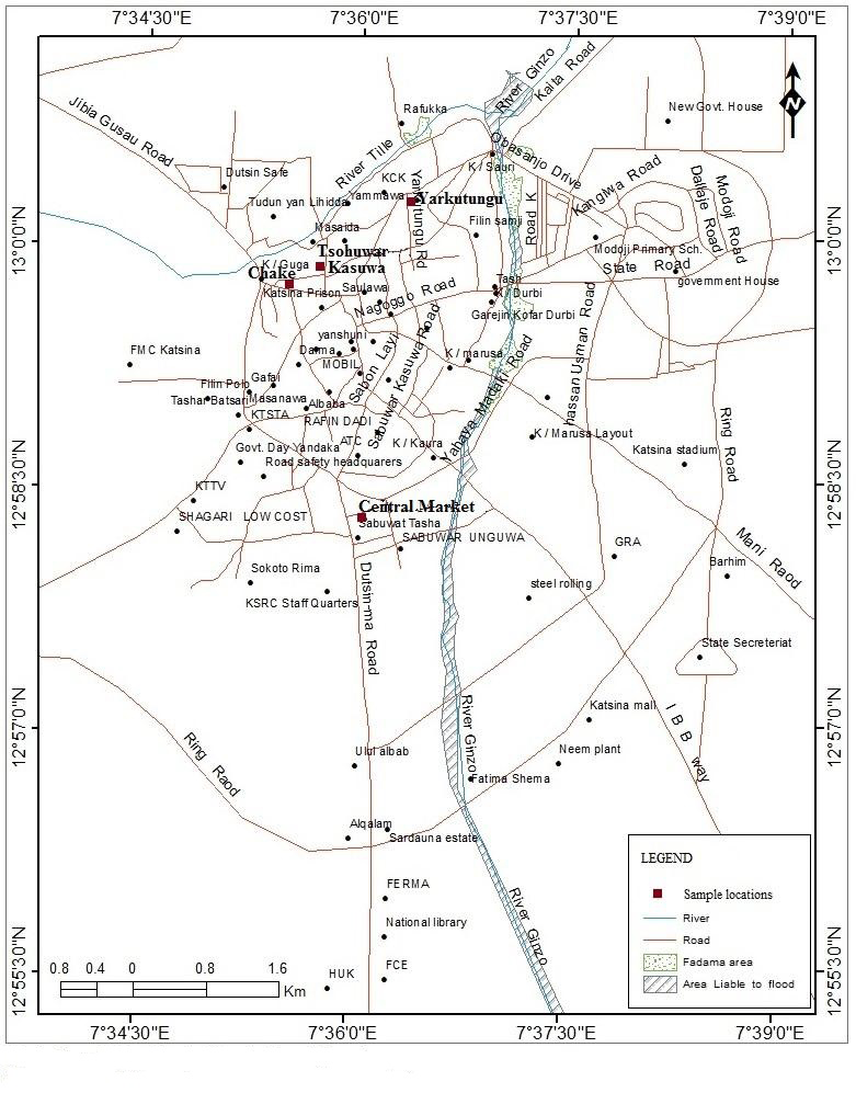
Sampling was carried out in Central, Yar Kutungu, Chake and Tsohuwar Kasuwa markets of Katsina metropolis, Katsina State. Katsina is a state in the northwestern geopolitical zone of Nigeria. According to National population commission (NPC, 2006), Katsina’s estimated population was 5, 792, 578 people. The state is bounded by Niger republic to the north, Jigawa and Kano states to the east, Kaduna state to the south and Zamfara state to the west Figure 1.
Sample Collection
A total of Sixteen (16) samples (Four each) of rice, wheat, maize and millet were collected from four selected major markets (Figure 1) and were transported to the laboratory of the Department of Biological Sciences, Al-Qalam University Katsina for subsequent analyses.
Sample Processing
About 80% of the collected samples were grounded to powdered form using sterilized mortar and pestle, while the remaining 20% were kept for direct plating during fungal isolation. The samples were stored at 4oC pending further analyses [14].
Isolation and Identification of Fungi
Fungal isolation was carried out by direct plating method described by Gautam AK, et al. [15] and adopted by Shamsudeen, et al. [16]. Potato Dextrose Agar (PDA) with chloramphenicol (500mg/l) was prepared following the manufacturer’s instructions and sterilized by autoclaving at 121oC for 15 minutes, and allowed to cool. After cooling, prepared the medium was dispensed into petri-dishes and samples were inoculated onto the petridishes containing the prepared medium and incubated at room temperature for 5-7 days. After incubation, fungal colonies were purified by sub culturing onto freshly prepared PDA plates and incubated at room temperature for 5-7 days. Identification of pure fungal isolates was carried out by observing cultural characteristeristics such as pattern of growth and pigment production as well as microscopic characteristics such as hyphal morphology and conidia as described by the El- Hassan, et al. [17].
Microscopic Slide Preparation and Observation
A drop of mounting fluid, lacto phenol cotton blue solution was placed on a grease free slide. Scrapings of the pure isolates were taken from the Potato Dextrose Agar (PDA) and transferred on the fluid using a sterilized, cooled wire needle. It was then pressed gently to enable it mix properly with the stain. A sterile forcep was used to place a cover slip over the slides and blotting paper was also used to wipe excess stain and then examined under low magnification (x10) and high magnification (x40) objectives [17, 18].
Extraction of the Samples
Thirty (30g) of the samples each was grounded using pestle and mortar. Twenty (20g) of each of the grounded samples was measured into a clean conical flask with seal. A 100ml of 70:30(v/v) methanol-water solutions was added and the flasks were sealed. The mixture was vigorously shaken for three (3) minutes, allowed to settled and then filtered through a filter paper and the filtrate was obtained, the residue was discarded and the filtrate was concentrated using a rotary evaporating machine. The concentrated extracts were poured in clean bijou bottles, labeled and kept in a refrigerator for further analysis [19].
Detection of Aflatoxin in the Samples’ Extracts
The chromate graphic plates were coated with silica gel. Thirty grams (30g) of silica gel was mixed with 75ml of distilled water, this was then used to coat the chromatographic plates and allowed to dry for 30minutes. The coated plates were activated by heating in an oven at 100oC for30minutes, then, the extracts from extraction were spotted on the plates using capillary tubes. Chloroform: methanol mixture of 97:3ml was used as the chromatographic solvent; it was run into the chromatogram or chromatographic tank to serve as the mobile phase of the chromatography. The spotted plates were dipped into the tank containing the solvent without allowing the solvent to touch the areas spotted with the extract. The solvent was allowed to move the substance (extracts) until the solvent stop moving. The distance moved by the solvent and that moved by the substance was marked immediately. the retention factor was determined using the relation; RF=Distance Moved by Substance (DMS)/Distance Moved by Solvent (SF) Aflatoxins was then detected by illuminating the plates with ultra violet light (UV) where blue or green fluorescence indicate aflatoxin B and G respectively [20].
Results
Identification of Fungal Isolates
The macroscopic and microscopic features of the identified fungal species were presented in (Table 1). The species identified were A. niger, A. flavus, A. fumigatus, and A. parasiticus.
| Macroscopic Features | Microscopic Features | Species Identified | |
|---|---|---|---|
| W | Black mycelia growth and fully extended in the growth medium | Conidiophores that are not branched with bulb like end bearing conidia | A. Niger |
| X | Dark green colony that are umbonate | Spherical conidia beared on rough conidiophore | A. parasiticus |
Table 1: Macroscopic and Microscopic Features of Fungi Species Identified
conidial heads were typically columnar. Conidio- phore stipes were short, smooth walled conidia globose to subglobose, A. fumigates
- Y
- Bluish green colony, that are hairy and flat
- Z
- Green colony with flat edges conidial heads were typically radiate, conidia were globose to subglobose,
- A. flavus
Table 2: Macroscopic and Microscopic Features of Fungi Species Identified
| Sampling Location | Samples | A. niger | A. flavus | A. parasiticus | A. fumigatus |
|---|---|---|---|---|---|
| Chake Market | MZ | - | - | + | - |
| RC | + | + | - | - | |
| ML | - | - | + | - | |
| WH | + | - | + | - | |
| Central Market | MZ | - | + | - | + |
| RC | + | - | - | - | |
| ML | + | - | - | - | |
| WH | + | - | - | - | |
| T/Kasuwa Market | MZ | - | - | + | - |
| RC | - | + | - | - | |
| ML | - | - | + | - | |
| WH | - | - | + | - | |
| Yarkutungu Market | MZ | + | - | - | - |
| RC | - | + | - | - | |
| ML | - | + | - | - | |
| WH | + | - | - | - |
Table 3: Fungal Species Isolated From the Samples
Key: MZ= Maize, RC = Rice, ML = Millet, WH = Wheat, + = Present, - = Absent Table 2: Fungal Species Isolated From the Samples
Occurrence of Fungal Species in the Samples
The occurrence of fungal species was presented in table 3. A parasiticus had the highest occurrence of 6(35.3%) followed by A_. flavus_ and A. niger with the occurrence of 5(29.4%) each. the lowest of occurrence of 1(5.9%)was recorded with A. fumigatus.
| Species | Occurrence | Percentage Occurrence (%) |
|---|---|---|
| A. niger | 5 | 29.4 |
| A. flavus | 5 | 29.4 |
| A. parasiticus | 6 | 35.3 |
| A. fumigates | 1 | 5.9 |
| Total | 17 | 100 |
Table 4: Occurrence of Fungal Species in the Samples
Colour of the Samples’ Extracts
The colour of the samples extracts collected from various markets was presented in Table 4. The colours are dark yellow, yellow, pale yellow, turbid white, white and dark brown.
| Sampling Location | Samples | Colour of the Extracts |
|---|---|---|
| Chake Market | MZ | Dark Yellow |
| RC | White | |
| ML | Pale Yellow | |
| WH | White | |
| Central Market | MZ | Yellow |
| RC | Yellow | |
| ML | White | |
| WH | Turbid White | |
| Tsohuwar Kasuwa Market | MZ | Pale Yellow |
| RC | White | |
| ML | Pale Yellow | |
| WH | Pale Yellow | |
| Yarkutungu Market | MZ | Yellow |
| RC | White | |
| ML | Yellow | |
| WH | Dark Brown |
Table 5: Colour of the Samples Extracts
Key: MZ= Maize, RC = Rice, ML = Millet, WH = Wheat Table 4: Colour of the Samples Extracts
Retention factor, Florescence and Aflatoxin Detected in the Samples
Table 5 shows the retention factor and aflatoxin detected in samples. The distance moved by the solvent (DMS) ranges from 2.4-2.6cm and solvent front (SF) ranges from 1.3 to 2.3 cm. while the RF ranges from 0.52 to 1.9. Blue florescence (i.e aflatoxin B) was detected in samples collected from Chake, Yarkutungu and Tsohuwar Kasuwa markets but was not detected in samples collected from Central Market (Table 5).
| Samples | DMS(cm) | SF (cm) | RF | Florescence | Aflatoxin Detected | |
|---|---|---|---|---|---|---|
| Chake Market | MZ | 2.4 | 1.9 | 0.76 | Blue | AFB |
| Chake Market | RC | 2.4 | 1.4 | 0.56 | Blue | AFB |
| Chake Market | ML | 2.4 | 1.8 | 0.75 | Blue | AFB |
| Chake Market | WH | 2.4 | 1.9 | 0.76 | Blue | AFB |
| Central Market | MZ | 2.6 | 1.8 | 1.9 | ND | Absent |
| Central Market | RC | 2.6 | 2.3 | 1.4 | ND | Absent |
| Central Market | ML | 2.6 | 1.9 | 1.8 | ND | Absent |
| Central Market | WH | 2.6 | 2 | 1.9 | ND | Absent |
| Tsohuwar Kasuwa Market | MZ | 2.5 | 2.1 | 0.88 | Blue | AFB |
| Tsohuwar Kasuwa Market | RC | 2.5 | 1.9 | 0.79 | Blue | AFB |
| Tsohuwar Kasuwa Market | ML | 2.5 | 1.9 | 0.79 | Blue | AFB |
| Tsohuwar Kasuwa Market | WH | 2.5 | 1.3 | 0.54 | Blue | AFB |
| Yarkutungu Market | MZ | 2.5 | 1.8 | 0.72 | Blue | AFB |
| Yarkutungu Market | RC | 2.5 | 1.8 | 0.72 | Blue | AFB |
| Yarkutungu Market | ML | 2.5 | 1.3 | 0.52 | Blue | AFB |
| Yarkutungu Market | WH | 2.5 | 1.7 | 0.62 | Blue | AFB |
Table 6: Retention Factor, Florescence and Aflatoxin Detected
Key: DMS = Distance Moved by the Solvent, SF = Solvent Front, RF = Retention Factor, AFB = Aflatoxin B, ND = Not Detected. Table 5: Retention Factor, Florescence and Aflatoxin Detected
Discussion
The present study revealed that samples collected from major markets of Katsina Metropolis were contaminated by .A. niger, A. flavus, A. parasiticus, and A. fumigatus. The presence of these fungal species is an indication of possible health hazard as some species of the genus Aspergillus were reported to cause food intoxication and poisoning through production of toxic secondary metabolites termed aflatoxins [21]. Fungal contamination of food is a major concern in food safety as huge quantities of foods are wasted annually, because this contamination renders the food unfit for human and animals consumption as a result of physical damage and reduction of its nutritional value [7]. Also, fungal food spoilage and toxicities have resulted in illnesses and deaths of humans, animals and plants as well as the spoilage of large percentage of food products leading to significant decline in plants and animals productivities and overall economic loss in many regions of the world [22, 23, 24].
The occurrence of these species in cereal grains was reported by many researchers [17, 20, 25]. The presence of these species in the samples could be due to careless and improper handling, poor storage and unhygienic conditions as well as favorable environmental factors that favor the growth of food spoilage fungi [25].
In this study, analysis of samples’ extracts using thin layer chromatography revealed the retention factor (RF) values that ranged from 0.52-1.9. Florescence was not detected in samples with retention factor value above 0.88. Blue florescent which indicated aflatoxin B was detected in samples with retention factor of 0.52-0.88. These retention factors were within the ranges to which aflatoxin B was detected by El- Hassan, et al. [17, 20, 26, 27] reported that when similar compounds are extracted using the same method, and developed on thin layer chromatography using the same stationary and mobile phases, they are likely to have the same retention factor values. Occurrence of aflatoxin in any sample due to contamination by aflatoxigenic fungi either in field or during storage, makes the sample unfit for human and animal consumption as aflatoxins are group of toxic and carcinogenic secondary metabolites of fungal origin. A. flavus, A. parasiticus, and rarely other species and strains of Aspergillus were reported as potent aflatoxin producers [28]. Aflatoxins are very powerful hepatocarcinagens and their naturally occurring mixtures have been classified as a class I human carcinogen [29]. They play role in causing liver damage, liver cirrhosis and induction of tumor in both man and animals [16]. Health problems such as infertility in men [30] and reduction in salivary secretory immunoglobulin A levels [31] were reported.
Conclusion
The results of this study showed that cereal samples analysed were contaminated with one or more fungal species that are known to cause food poisoning and foodborne intoxication. Detection of aflatoxin B in some samples is a potential health hazard as aflatoxins were reported to induce cancers, tumors and other health problems. Therefore, fungal and aflatoxin contaminations of cereals at the field, during harvest or storage should be controlled through good agricultural processing and storage practices as well as ensuring hygienic practices at the point of sales.
References
-
Mohammed EZ (2011) Impact of Mycotoxins on Humans and Animal. Journal OF Saudi Chemical Society, 15(2): 129-144.
-
Shamsuddeen U, Ahmad MA, Abdulkadir RS (2017) Evaluation of aflatoxin contamination in Zeamays (maize)soldi Katsina central market, Nigeria.UMYU J Microb Resear2(1):102-106.
-
Pitt J I, Wild CP, Gelderblom W, Miller J, Riley RT, et al. (2012) Improving public health through mycotoxin control (Publication No. 158) International Agency for Research on Cancer 1-2.
-
Ashiq S, Hussain B, Ahmad B (2014) Natural occurrence of mycotoxins in medicinal plants: A review. Article in Fungal Genetics and Biology 66(1): 1-10.
-
Bennet JW, Klich M (2003) Mycotoxins. Clinical Microbiology Review 16: 497-516.
-
Wambacq E, Vanhoutte I, Audernaert K, De Gelder L, Haesaert G (2016) Occurrence, Prevention and Remediation of Toxigenic Fungi and Mycotoxins in Silage: A Review. J Sci Food Agri 96: 2284-2302.
-
Sule E I, Umoh UJ, Whong CMZ, Abdullahi IO (2014) Aflatoxin-producing ability of fungal isolates from maize and maize products. Biol environ sci j tropics 11(2): 33- 36.
-
Wu F (2007) Measuring the economic impacts of Fusarium toxins in animal feeds. Animal Feed Science and Technology 137(3): 363374.
-
Kumar P, Mahato D K, Kamle M, Mohanta T K, Kang S G (2017). Aflatoxins: A global concern for food safety, human health, and their management. Front Microbial 7(468): 1-10.
-
Navale V, Dhuri V (2021) Aspergillus Derived Mycotoxin in Food and the Environment: Prevalence, Detection and Toxicity. Toxicology Reports 8: 1008-1030.
-
Ellis WO, Smith JP, Simpson BK (1991) Aflatoxins in food occurrence, biosynthesis, effects on organisms, detection and methods of control. Critical Review on Food Science and Nutrition 30(4): 403-439.
-
Murphy PA, Hendrich S, Landgren C, Bryant CM (2006) Food mycotoxins: an update. Journal of Food Science 71:R51-R65.
-
Mahato DK, Lee KE, Kamle M, Devi S, Krishna ND, et al. (2019) Aflatoxins in Food and Feed: An Overview on Prevalence, Detection and Control Strategies. Frontiers in Microbiology 10: 2266.
-
Sadhasivam S, Britzi M, Zakin V, Kostyukovsky M, Tronstanetsky A, et al. (2017) Rapid detection and identification of mycotoxigenic fungi and mycotoxins in stored wheat grain. Article Toxins 9(302): 1-17.
-
Gautam AK, Gupta H, Soni Y (2012) Screening of fungi and mycotoxins associated with stored rice grains in Himachal Pradesh. International Journal of Theoretical and Applied Sciences; 4(2): 128-133.
-
Shamsuddeen U, Magashi M A (2004) Determination of aflatoxin concentration in yellow and white maize. Biol environ j tropics 1(12): 91-94.
-
El Hassan FI, Edigba BA, Sadisu FU (2022) Mycoflora and Aflatoxin Producing Fungi from Some Storage Cereals Grains Sold in Darki Market, Wudil Local Government Area, Kano state, Nigeria. Asian Food Science Journal 21(1): 65-76.
-
Okafor SE, Eni AO (2018) Microbial quality and the occurrence of aflatoxins in plantain/yam and wheat flours in Ado-Odo Ota. IOP Conf Ser: Earth Environ Sci pp: 210.
-
Tijjani MB, Zango UU, Wada Kura A, Hosea HD (2013) Screening and quantification of aflatoxins present in sorghum obtained from open markets in Zaria. Bio Environ Sci J Trop 10 (2): 206-210.
-
Shamsuddeen U, Kabir A (2015) Study on aflatoxin contents of maize from Dawanau grain market in Kano, Nigeria. Paper Published in the 38th annual general meeting and scientific conference of Nigerian Society for Microbiology, Book of Abstract (pp-163). Lagos, Nigeria.
-
Keta N, Jibrin A, Majlinda, Joseph GG (2019) Incidence of Fungal flora and Aflatoxin content of Millet and Maize cereal grains sold in Guinea Savanna Zones of Kebbi State. Science World Journal 14(2): 12-15.
-
Patial V, Asrani RK, Thakur M (2018) Food-borne mycotoxicoses: pathologies and public health impact. in: foodborne diseases. Elsevier 239-274.
-
Singh P, Cotty PJ (2019) Characterization of Aspergilli from dried red chilies (Capsicum spp.): Insights into the etiology of aflatoxin contamination. Int J Food Microbiol 289: 145-153.
-
Salisu BD, Almajir IR (2020) Aflatoxins and aflatoxigenic fungal contamination of common poultry feed products in Katsina State, Nigeria. Nov Res Microbiol J 4(1): 653- 665.
-
Salisu DB, Siti MA, Wan WRI, Mazlan N (2020) Incidence, Distribution and Phenotypic Characterization of Fungi Contaminating Commonly Consumed Food Grains in Nigeria. Malaysian Jounal of Medicine and Health Sciences, 16(11): 18-27.
-
Gurav NP, Medhe S (2018) Analysis of Aflatoxins B1, B2, G1 and G2 in Peanuts: Validation Study. Anal Chem Ind J 17(2):126.
-
Matome G, Hunja M, Akebe LK, Michael P (2017) Morphological characterization and determination of aflatoxin-production potentials of Aspergillus flavus isolated from maize and soil in Kenya. Agriculture 7(10): 80-94.
-
Mazaheri M (2009) Determination of aflatoxins in imported rice to Iran. Food Chemical Toxicology, 47(8): 2064-2066.
-
International Agency for Research on Cancer (IARC) (1993) IARC Monographs on the Evaluation of Carcinogenic Risks to Humans, Some naturally occurring substances: food items and constituents, heterocyclic aromatic amines and mycotoxins. IARC Monogr Evaluation of Carcinogenic Risks in Human 56: 489-521.
-
Uriah N, Ibeh IN, Oluwafemi F (2001) A study on the impact of aflatoxin on human reproduction. Advance Journal of Reproductive. Health, 5(1): 106-110.
-
Turner PC, Moore SE, Hall A J, Prentice AM, Wild CP (2003) Modification of immune function through exposure to dietary aflatoxin in Gambian children. Environmental Health Perspective 111(2): 217-220.
- Diversity of Candida sp and Antifungal Susceptibility Patterns in Digestive Candidiasis among People Living with HIV in CHU of Libreville, Gabon
- Vulvovaginal candidiasis: Retrospective study (2019- 2021) at the Centre Hospitalier National de Pikine, Suburban Dakar, Senegal
- Identification of Environmental Fungal Species in Clinical Services of University Hospital of Angre, Abidjan (Cote d’Ivoire)
- New Location of some Gasteroid Basidiomycetes in Western Kazakhstan
- Evaluation of Various Extracellular Enzymes of Ectomycorrhizal Mushrooms
- Morphology and Phylogeny of Lactarius Wallichianae sp. nov and Xerula magnispora sp. nov. from India