Resolution of Bilateral Exudative Retinal Detachment Secondary to Renal Hypertension with Systemic Management
Exudative retinal detachment (ERD) is an ocular condition, which develops from pathological conditions that disrupt the integrity of blood-retinal barrier due to fluid accumulation in sub retinal space or under neurosensory retina. Exudative retinal detachment is typically associated with inflammatory, infectious, neoplastic, and vascular pathological conditions. We report the management of bilateral exudative retinal detachment in young patients secondary to Renal Hypertension.
Introduction
Exudative retinal detachment (ERD) is a type of retinal detachment that develops from pathological conditions that disrupt the integrity of the blood-retinal barrier and cause fluid accumulation in sub retinal space. If not treated on time, it can damage the retinal photoreceptors, which can cause irreversible blindness.
Case Report
A 19-year-old male presented to us complaining of severely diminished vision in both eyes for one week associated with headache and vomiting. The patient has had a history of paraplegia since birth following Meningomyelocele surgery, which also resulted in a neurogenic bladder with grade IV Vesicoureteral reflux. The BCVA and IOP in the right and left eyes were 20/1200 and 20/200, and 12 and 14 mmHg respectively. Dilated fundus examination of both eyes revealed multiple peripapillary white patches appearing like cotton wool spots around the disc with mild blurring of disc margin, a few superficial hemorrhages, areas of sub retinal folds and fluid inferiorly suggestive of serous retinal detachment involving the macula; these findings corresponded to OCT imaging (Figure 1a, 1b, 1c, 1d).
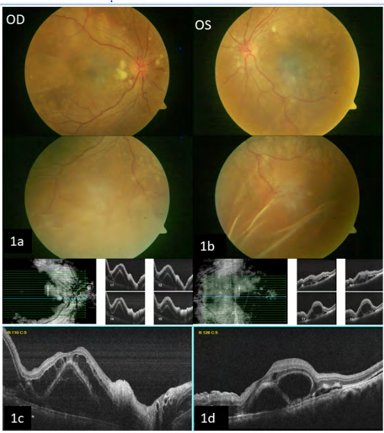
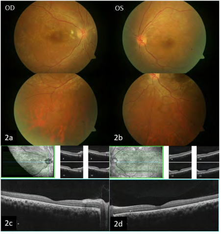
No evident break was noted in the periphery. The B scan showed a shifting fluid sign bilaterally. Hence, he was diagnosed with bilateral exudative retinal detachment (ERD) and was urgently referred to a physician and nephrologist for systemic evaluation. The recorded blood pressure was 200/120 mmHg, and B scan KUB showed bilateral hydronephrosis. The patient was started on Anti- hypertensive medications (Prazosin 5 mg OD, Telmisartan 40 mg OD, Cilnidipine 10 mg BD) On 1-month follow-up post-treatment, the visual acuity improved to 20/40 (6/12) in both eyes. Fundus examination showed a near total resolution of sub retinal fluid and resolving cotton wool spots and hemorrhages, supported by the OCT (Figure 2a, 2b, 2c and 2d).
Discussion
Exudative retinal detachment is typically associated with inflammatory, infectious, neoplastic and vascular pathological conditions [1, 2]. A similar case was noted by Otuka et al., where they managed the exudative detachment with systemic hypertension control and hemodialysis [3]. Blood pressure control and the balance of fluids are of prime importance in patients with renal failure, and a multidisciplinary approach is warranted. It is extremely important to understand the underlying pathology of retinal detachment. Our group recommends ophthalmic evaluation for all chronic kidney patients to prevent irreversible loss of vision.
Conclusion
With early recognition of the underlying cause, ERD can resolve with a good visual prognosis. A multidisciplinary approach is the key.
Financial Support and Sponsorship
Nil.
Conflicts of Interest
There are no conflicts of interest.
Declaration of Patient Consent
The authors certify that they have obtained all appropriate patient consent forms. In the form the patient(s) has given his consent for his images and other clinical information to be reported in the journal. The patients understand that their names and initials will not be published in any form.
References
-
Amer R, Nalci H, Yalcindag N (2017) Exudative retinal detachment. Surv Ophthalmol 62(6): 723-769.
-
Ghazi NG, Green WR (2002) Pathology and pathogenesis of retinal detachment. Eye (Lond) 16(4): 411-421.
-
Otuka OAI, Eweputanna LI, Okoronkwo NC, Kalu A (2021) Bilateral Exudative Retinal Detachment in a Young Patient with Chronic Renal Failure. Int Med Case Rep J 14: 139-144.
- Screening of Hospital Staff During World Glaucoma Week in a Tertiary Eye Care Centre
- Angioid Streaks with Macular Neovascularization: Clinical Insights from Two Cases
- Giant Kissing Naevus: An Oculoplastic Challenge
- Why Freedom of Vision Should Not Cost the Freedom of Feeling - LASIK in the Climate of Change
- Asymmetric Optic Nerve with Small Disc and Large Cup: A Rare and Challenging Case of Unilateral Optic Nerve Hypoplasia
- Large Angle Exotropia in a Child: A Case Report