Multimodal Imaging of Peripheral Combined Hamartoma of Retina and Retinal Pigment Epithelium
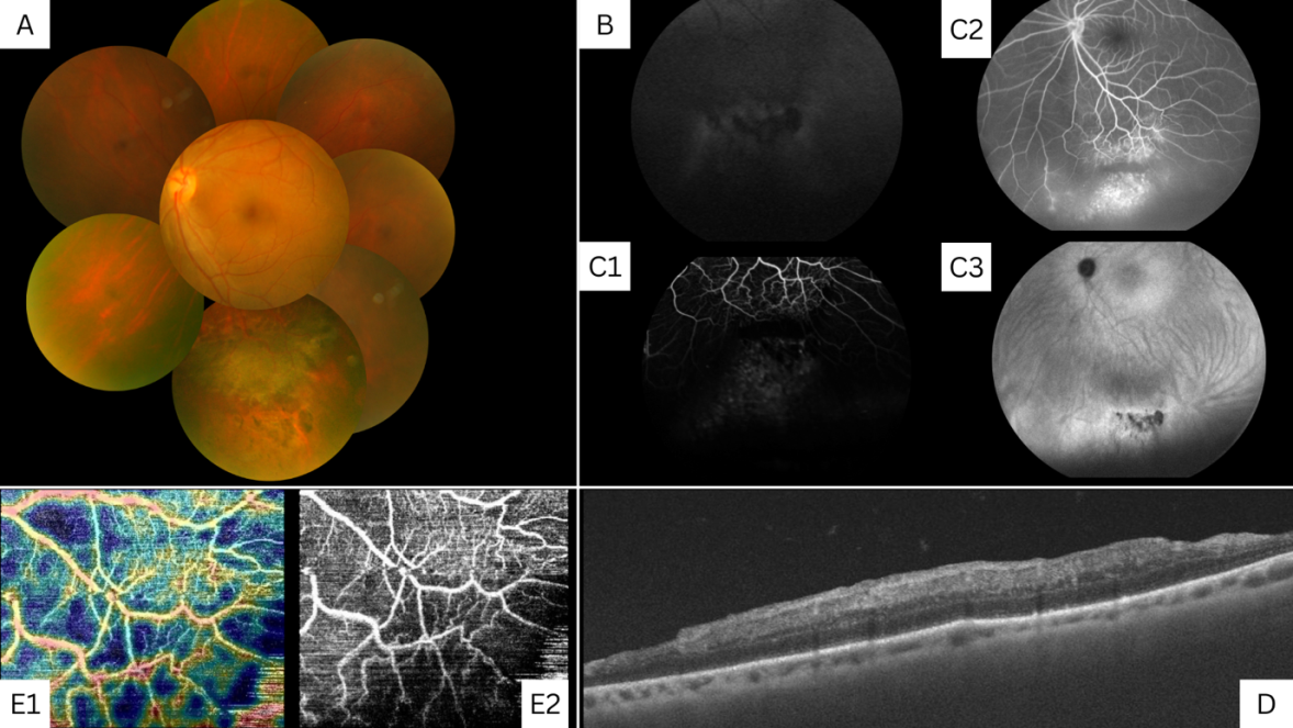
A 25-year-old female presented to our clinic for a routine eye examination. Her best-corrected visual acuity was 6/6, N6 in both eyes. Anterior segment examination of the right eye was unremarkable, and the left eye showed conjunctival melanosis in the supero-temporal quadrant. On fundus examination, the right eye was within normal limits; the left eye showed a flat pigmented lesion with distinct margins, tortuosity of the overlying vascular architecture, and surrounding RPE alterations in the infero-temporal quadrant, as shown in the colour fundus photograph. In this clinical imaging, we highlight the multimodal imaging of CHRRPE, which is rarely found peripherally (Figure 1).
Image Article
A 25-year-old female presented to our clinic for a routine eye examination. Her best-corrected visual acuity was 6/6, N6 in both eyes. Anterior segment examination of the right eye was unremarkable, and the left eye showed conjunctival melanosis in the supero-temporal quadrant. On fundus examination, the right eye was within normal limits;
Image Article
the left eye showed a flat pigmented lesion with distinct margins, tortuosity of the overlying vascular architecture, and surrounding RPE alterations in the infero-temporal quadrant, as shown in the colour fundus photograph. In this clinical imaging, we highlight the multimodal imaging of CHRRPE, which is rarely found peripherally (Figure 1).
Conflict of Interest
The authors have no competing financial interests to disclose and no funding was received from any external sources.
- Screening of Hospital Staff During World Glaucoma Week in a Tertiary Eye Care Centre
- Angioid Streaks with Macular Neovascularization: Clinical Insights from Two Cases
- Giant Kissing Naevus: An Oculoplastic Challenge
- Why Freedom of Vision Should Not Cost the Freedom of Feeling - LASIK in the Climate of Change
- Asymmetric Optic Nerve with Small Disc and Large Cup: A Rare and Challenging Case of Unilateral Optic Nerve Hypoplasia
- Large Angle Exotropia in a Child: A Case Report