Management of Optic Disc Pit Associated Neurosensory Detachment
A 22 year old male presented to us with complaints of seeing wavy lines in right eye of 3 days duration. He also gave history of reduced vision in left eye since childhood. His best corrected visual acuity was 6/18, N18 and 6/24, N12 in right and left eye respectively.
Image Article
A 22 year old male presented to us with complaints of seeing wavy lines in right eye of 3 days duration.
Image Article
He also gave history of reduced vision in left eye since childhood. His best corrected visual acuity was 6/18, N18 and 6/24, N12 in right and left eye respectively.
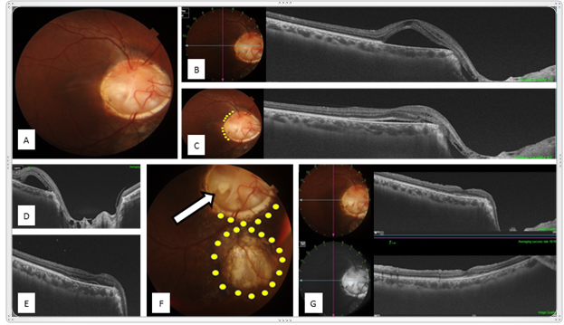
a circumferential pattern at the temporal edge of the disc, following which resolution of the NSD was noted. Patient was doing well for 2 weeks, following which he presented to us again with sudden onset of metamorphopsia of 1 day duration with neurosensory detachment at fovea. Patient was planned for pars plana vitrectomy with scleral patch graft at the optic disc in order to plug the defect, following which resolution of the NSD was noted on day 1 and sustained up to 1 month, with improvement in best corrected visual acuity to 6/6P in snellen chart. Patient presented to us again with metamorphopsia and sudden vision blur in right eye at 40 days post surgery, with drop in visual acuity to 6/12. Neurosensory detachment was noted again at the fovea with apparently well taken scleral graft at the optic disc. We were unable to ascertain the source of neurosensory fluid, and a presumptive source of fluid origin from the coloboma edge was made. Barrage laser was performed in circumferential pattern around the coloboma edges as well as inferior edge of optic disc. At 6 months follow up, no evidence of sub- retinal fluid was noted with maintenance of visual acuity of 6/6P. In this article we highlight the importance of ruling out the source of fluid in order to prevent recurrences.
- Screening of Hospital Staff During World Glaucoma Week in a Tertiary Eye Care Centre
- Angioid Streaks with Macular Neovascularization: Clinical Insights from Two Cases
- Giant Kissing Naevus: An Oculoplastic Challenge
- Why Freedom of Vision Should Not Cost the Freedom of Feeling - LASIK in the Climate of Change
- Asymmetric Optic Nerve with Small Disc and Large Cup: A Rare and Challenging Case of Unilateral Optic Nerve Hypoplasia
- Large Angle Exotropia in a Child: A Case Report