Granular Cell Tumor, When to Think About it? Clinical Case
We present the case of a female patient with a lesion on the dorsal of the tongue, of two years of evolution, whose histopathological diagnosis was granular cell tumor. We are recapitulating the epidemiological, clinical, histological, treatment and prognosis characteristics, for a better understanding of the pathology
Clinical Case
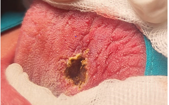
A 47-year-old female presented with a nodular lesion on the dorsal medial side of the tongue, approximately 1 cm, circular, hard, slightly painful, with defined margins, papillomatous in appearance, with a 2-year history.
She underwent surgical resection and the pathology report was: granular cell tumor, with associated pseudoepitheliomatous hyperplasia, 9 x 9 mm, with lesion- free surgical edges.
The surgical site is shown, after resection (Figure 1). The patient is asymptomatic to date.
It is a rare tumor, with a predilection in females, between the fourth and sixth decade of life, although cases have been reported in childhood [1]. It was described by Abrikossoff in 1926. It occurs mainly in the oral mucosa, skin and subcutaneous cellular tissue, but can be located in other organs (breast, thyroid, mediastinum, respiratory and gastrointestinal tract, biliary tree, pancreas, ovary-testis, urinary system, heart and central nervous system) [2], its malignant degeneration is rare (it is accepted only if it has produced metastasis, which happens in 2-2.5% of cases and with the histological presence of 3 or more of the following criteria : necrosis, spindle cell morphology, vesicular nucleus with prominent nucleolus, more than 2 mitoses per 10 high- power fields, elevated nucleo-cytoplasmic index, and cellular pleomorphism) [3]. It is generally unique, although there are multiple cases, especially in childhood, related to Noonan Syndrome (rasopathy, relatively frequent, characterized by presenting a genetic mutation in the encoding of Ras family proteins, affecting the cell proliferation and differentiation, and characterized by short stature, congenital heart disease, and a characteristic facial phenotype) and with neurofibromatosis [4].
Thanks to advances in immunohistochemistry, it is believed that its histological origin is Schawnn cells [1].
Histologically, tumor cells often show large eosinophilic granules surrounded by a transparent halo, known as pustule-ovoid bodies of Milian. The overlying epithelium often shows prominent pseudoepitheliomatous hyperplasia that can be confused with squamous cell carcinoma if the biopsy is superficial. Immunohistochemistry is positive for S100 protein, CD68 antigen, and neuron-specific enolase [5].
Simple excision is the treatment of choice for benign presentation, but local recurrences can occur, which are more frequent in cases with positive surgical margins, but local recurrences have also been reported in tumors excised with free margins, so it is recommended a follow-up of several years [6].
Conclusion
Despite the fact that the granular cell tumor is an infrequent pathology, we must bear in mind these diagnostic possibilities when dealing with lesions with similar characteristics and not think only of papillomas of the oral cavity.
References
-
Machado I, Cruz J, Lavernia J, Llombart-Bosch A (2016) Solitary, multiple, benign, atypical, or malignant: the “Granular Cell Tumor” puzzle. Virchows Arch 468(5): 527-538.
-
Calonje E, Tsai KE (2016) Soft-tissue tumours and tumour-like conditions. Chapter 137 Griffiths CEM. In: Barker J, Bleiker TO, et al. (Eds.), Rook’s Textbook of Dermatology. 9th (Edn.), Wiley, London, UK.
-
Fanburg JC, Meis-Kindblom JM, Fante R, Kindblom LG (1998) Malignant granular cell tumor of soft tissue: diagnostic criteria and clinicopathologic correlation. The American journal of surgical pathology 22(7): 779-794.
-
Muscardin LM, Paradisi M, Provini A, Cota C, Marzetti G (2006) Multiple cutaneous granular cell tumors, joint hypermobility and mild facial dysmorphism in a child. International Journal of Dermatology 45(7): 847-850.
-
Torrijos-Aguilar A, Miquel VA, Pitarch-Bort G, Garcia PM, Fortea-Baixauli JM (2009) Cutaneous granular cell tumor: a clinical and pathologic analysis of 34 cases. Actas dermo-sifiliograficas 100(2): 126-132.
-
Mirza FN, Tuggle CT, Zogg CK, Mirza HN, Narayan D (2018) Epidemiology of malignant cutaneous granular cell tumors: A US population-based cohort analysis using the Surveillance, Epidemiology, and End Results (SEER) database. Journal of the American Academy of Dermatology 78(3): 490-497.
- 4th Branchial Cleft Sinus Anomaly Presenting as Recurrent Thyroid Abscess in A Child: A Case Report
- Parotid Duct Injury Repaired Using an Angiocatheter Stent: A Case Report
- Organization and Functionality of the Referral and Counter-Referral System for ENT Disorders in District Hospitals of N’Djamena, Chad: A Cross-Sectional Analytical Study
- Facial Metastases from a Gastrointestinal Stromal Tumor: A Case Report
- Panorama of Ent Cancers and Literature Review: Epidemiological Profile and Therapeutic Management
- Could Antimicrobial Resistance Prove to Be Both a Threat and an Opportunity for us?