Angiosarcoma of the Nose- A Rare Case Report Emphasizing Atypical Site and Literature Review
Angiosarcoma is a rare and aggressive malignant tumor primarily originating from blood and lymphatic vessels. While it commonly arises from cutaneous sites like the scalp, atypical presentations, such as nasal involvement, pose significant diagnostic challenges. This report presents the case of a 74-year-old male with a rapidly progressive violaceous mass on the nasal sidewall, diagnosed as angiosarcoma. The patient had no history of trauma, radiation, or lymphedema. Biopsy findings, along with immunohistochemical markers such as CD31, CD34, and D2-40, confirmed the diagnosis. The patient underwent subtotal rhinectomy with bilateral selective neck dissection, followed by adjuvant radiotherapy. Post-treatment follow-up showed no recurrence or metastatic disease. This case underscores the importance of considering angiosarcoma as a differential diagnosis in atypical sites and highlights the benefits of early diagnosis and multimodal treatment, which significantly improves survival rates. Further research is needed to establish clear guidelines for surgical margins and comprehensive treatment strategies.
Rockey Dahiya, M.B.B.S*, Natalie Weiss, M.D, Samip Patel, M.D and Phillip Pirgousis, M.D
Division of Head and Neck Surgery, Mayo Clinic, USA
Abbreviations
SEER: Surveillance Epidemiology and End Results; AJCC: American Joint Committee on Cancer Staging.
Introduction
Angiosarcoma is a rare, aggressive malignant tumor that commonly arises from blood and lymphatic vessels. It accounts for 15% of all head and neck sarcomas and approximately 2% of total soft tissue sarcomas [1]. Although it has the potential to arise from any cutaneous sites, it mostly involves the scalp.
Suspicion of angiosarcoma is primarily guided by pathological examinations, along with the physical examination findings such as location of the lesion, clinical history, and history of lymphedema or radiation exposure. Therefore, it can be difficult to promptly diagnose if it involves an atypical site like the nose, especially in patients without any history of risk factors such as exposure to causative agents, trauma, radiation, or lymphedema.
Despite its well-known severity and poor prognosis (5- year survival rate of 38%) [2], there is persistent uncertainty in considering it as a differential diagnosis for atypical sites. This case report illustrates the aggressive nature of angiosarcoma in an atypical site and highlights clues for its prompt diagnosis and surgical management.
Case Report
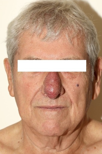
A 74-year-old male presented with a violaceous mass measuring 5.7 by 4.2 cm located on the entire nasal dorsum, which was progressive in nature. Eight weeks back, the patient noticed an erythematous lesion that within weeks, developed into a vascularized violaceous growth (Figure 1). The patient did not have any history of trauma, radiation, or lymphedema. The differentials considered include eczema, pyogenic granuloma, kaposi sarcoma, hemangioma, and angiosarcoma.
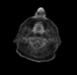
Figure1: Gross image showing vascularized violaceous growth on physical examination.
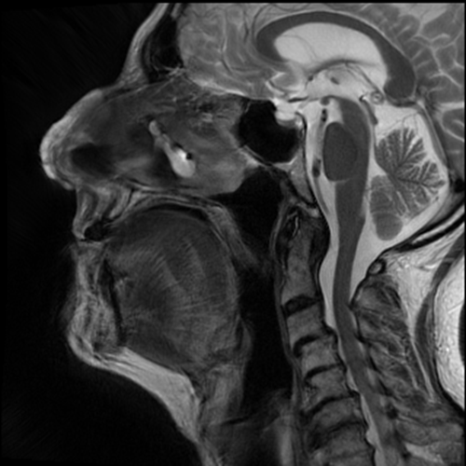
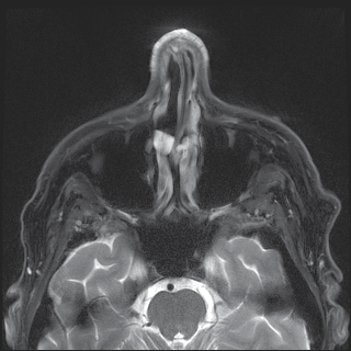
Biopsy demonstrated an atypical dermal stromal reaction and dilated vessels at the margins, raising suspicion of angiosarcoma. A PET scan revealed hypermetabolic ill- defined cutaneous and subcutaneous thickening of the nose, along with prominent level I b and IIa lymph nodes that were indeterminate for metastatic disease (Figure 2). Immunohistochemistry showed positivity for CD 31, CD 34, and D2-40. Additional MRI head and neck also revealed thickening and heterogenous enhancement of the entire soft tissue of the nose, with a small T2 hyperintense skin nodule on the anterior left cheek (Figures 3 & 4). The patient was promptly taken to the operating room for mapping biopsies to determine the extent of disease. Intraoperative sections confirmed negative margins.
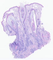
A formal surgical resection involving bilateral selective supra-omohyoid neck dissection was planned, which necessitating a subtotal rhinectomy (Figure 5). Surgical pathology revealed a unifocal cutaneous angiosarcoma of the nose of dimension, measuring 5.7 cm in greatest dimension. Histological examination indicated a high-grade sarcoma, exhibiting 16 mitosis figures, classified as stage pT4N0Mx. The harvested lymph nodes were negative for malignancy (Figure 6a & b). The patient had an uneventful postoperative course and was discharged within a week. Given lesion of more than 5cm, potential for subclinical disease in the bilateral neck and regional lymphatics, adjuvant radiotherapy was initiated, delivering a total dose of 6000 cGy in thirty fractions. Follow-up contrast enhanced CT scans at 1- and 3-month revealed no evidence of recurrent, residual, or metastatic disease (Figure 7). Subsequently, aesthetic reconstruction of the nasal dorsum was performed using a radial forearm free flap, along with split thickness harvested from thigh (Figure 5).
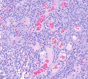
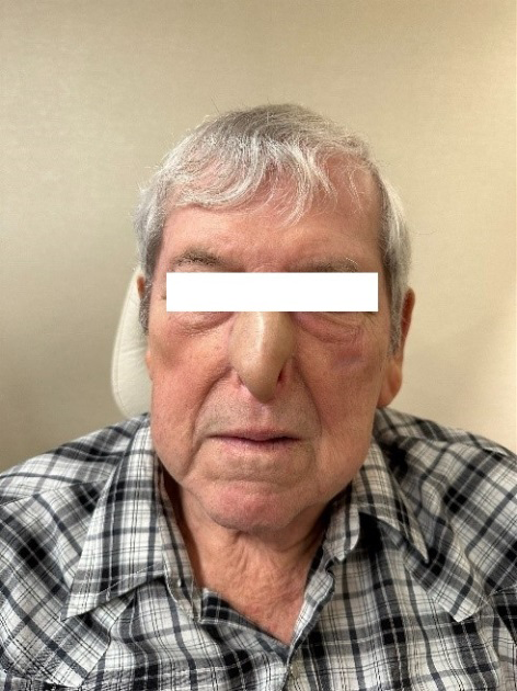
Figure 6a: H&E histological image of cutaneous angiosarcoma (20x).
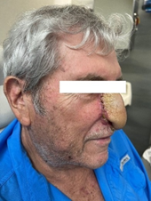
Figure 6b: Higher magnification of H&E of cutaneous angiosarcoma involving nose (40x) (shown in gross images), showing atypical dermal stromal reaction and dilated vessels in peripheral margins.
Discussion
In this report, we present an interesting case of angiosarcoma of the nose which initially presented as an erythematous lesion that rapidly progressed to a violaceous mass within a few weeks. It was managed surgically, followed by adjuvant radiation therapy. We aim to emphasize the importance of atypical site, aggressive biologic behavior, and early diagnostic findings for optimal treatment outcomes.
Between 1973 and 2007, the Surveillance, Epidemiology, and End Results (SEER) database reported involvement of the head and neck area in approximately 72% of patients diagnosed with cutaneous angiosarcomas [3]. Typically affecting older individuals (65-70 years) with a male- female ratio of 2:1. On gross examination, these lesions can present as blue or purple macular, nodular, plaque- like lesion with indistinct borders, multi-focal lesions, with or without lymphadenopathy over a short period. Prior reports have shown similarities on clinical examination between angiosarcoma and other conditions such as contact dermatitis, sebaceous cysts, rosacea, rhinophyma, and hemangioma. Overlapping gross characteristics in the early stage can lead to misdiagnosis of angiosarcoma at atypical site and consequently, use of unnecessary procedures and investigations. Therefore, prompt microscopic and immunohistochemical investigations are critical for accurate diagnosis. Additionally, the rapid growth pattern is also consistent with the diagnosis [4]. It is crucial to consider indeterminate features of any etiology at this stage. Early lesions with nonclassical phenotypic appearance should raise suspicion and repeat biopsies should be performed when diagnosis is uncertain.
Histopathologically, angiosarcoma can be categorized into several types such as lymphatic, vascular, and mixed types. Pathological criteria for diagnosis of vascular angiosarcoma include intravascular layering of atypical endothelial tumor cells, hyperchromasia, prominent irregular vascular spaces with a high proliferative index with mitotic figures. Early-stage angiosarcomas may be difficult to diagnose due to their subtle histologic features. Supporting the previous statement, Gravvanis et el. highlighted diagnostic difficulty, noting that the histological similarities between angiosarcoma and rhinophyma, particularly the presence of histiocytic cells with abundant cytoplasm, can complicate accurate diagnosis. Hence, a early comprehensive correlation of clinical and pathological findings with immunohistochemistry is necessary for accurate diagnosis [5]. Important histochemical markers include CD31, CD34, ERG, D2-40, and VEGF. However, lack of a consistent marker remains for differentiating angiosarcoma from other benign vascular proliferative diseases, as absence of D2-40 in tumor cells of angiosarcoma has been shown.
Wide surgical excision undoubtedly remains the most effective treatment for angiosarcoma. Nonetheless, the infiltrative nature of angiosarcoma often complicates the achievement of complete resection with clear margins, typically necessitating margins of 3 cm or more. Despite well-documented infiltrative behavior of this malignancy, clear guidelines for adequate margin width are lacking. Patients with comorbidities, advanced age, or multifocal lesions, present additional surgical challenges where excision may not be feasible, prompting consideration for adjuvant therapies. As given in our patient, radiation can be considered with curative intent in cases with high-risk target volume including the primary site and regional lymphatic at risk for subclinical disease in the bilateral neck. Multimodal therapy- incorporating both surgery and radiotherapy has emerged as a critical prognostic factor for improving overall survival rates. A few case reports have highlighted successful management with surgery or radiotherapy alone, particularly in cases where surgery is contraindicated due to comorbidities [6]. However, large cohort studies provide more robust data, two-year overall survival of 46% (p<0.0001) with the combination of surgery and radiotherapy, compared to only 11% with surgery or radiotherapy alone [7].While factors predicting recurrence have not been extensively evaluated, Grundahl et al. reported that the stage of the primary tumor (American Joint Committee on Cancer Staging (AJCC)), and tumor site, could help in predicting disease recurrence. Five-year survival rates have steadily improved from 10% to 30% [8], largely attributed to the multimodal approaches to management.
Conclusion
This report highlights the aggressive behavior of angiosarcoma at an atypical site, particularly the nose. We also aim to highlight the diagnostic challenges posed by the lack of distinct histological features and common risk factors such as trauma, or a history of radiation. Through a combination of surgical intervention and adjuvant radiotherapy, we successfully managed this patient, with no evidence of recurrence at follow-up. This review emphasizes the importance of considering angiosarcoma as a differential diagnosis regardless of the site involved. Despite the well- documented infiltrative nature of angiosarcoma, multimodal therapy appears to improve overall survival rates. However, continued research and clear guidelines on surgical margins and multimodal treatment strategies are necessary to further improve patient outcomes.
Disclosures
No Disclosures.
Conflict of Interest
The authors declare no conflict of interest.
References
-
Wanebo HJ, Koness RJ, MacFarlane JK, Eilber FR, Byers RM, et al. (1992) Head and neck sarcoma: report of the Head and Neck Sarcoma Registry. Society of Head and Neck Surgeons Committee on Research. Head Neck 14(1): 1-7.
-
Ramakrishnan N, Mokhtari R, Charville GW, Bui N, Ganjoo K (2022) Cutaneous Angiosarcoma of the Head and Neck-A Retrospective Analysis of 47 Patients. Cancers 14(15): 3841.
-
Albores-Saavedra J, Schwartz AM, Henson DE, Kostun L, Hart A, et al. (2011) Cutaneous angiosarcoma. Analysis of 434 cases from the Surveillance, Epidemiology, and End Results Program, 1973-2007. Ann Diagn Pathol 15(2): 93-97.
-
Aguila LI, Sánchez JL (2003) Angiosarcoma of the face resembling rhinophyma. J Am Acad Dermatol 49(3): 530-531.
-
Shustef E, Kazlouskaya V, Prieto VG, Ivan D, Aung PP (2017) Cutaneous angiosarcoma: a current update. J Clin Pathol 70(11): 917-925.
-
Sanchez Forero RA, Jaramillo LF, Ramirez J (2018) Cutaneous angiosarcoma in the nasal region treated with radiotherapy: case report. Med Univ 59(3): 1-10.
-
Pawlik TM, Paulino AF, McGinn CJ, Baker LH, Cohen DS, et al. (2003) Cutaneous angiosarcoma of the scalp: a multidisciplinary approach. Cancer 98(8): 1716-1726.
-
Mendenhall WM, Mendenhall CM, Werning JW, Reith JD, Mendenhall NP (2006) Cutaneous angiosarcoma. Am J Clin Oncol 29(5): 524-528.
- 4th Branchial Cleft Sinus Anomaly Presenting as Recurrent Thyroid Abscess in A Child: A Case Report
- Parotid Duct Injury Repaired Using an Angiocatheter Stent: A Case Report
- Organization and Functionality of the Referral and Counter-Referral System for ENT Disorders in District Hospitals of N’Djamena, Chad: A Cross-Sectional Analytical Study
- Facial Metastases from a Gastrointestinal Stromal Tumor: A Case Report
- Panorama of Ent Cancers and Literature Review: Epidemiological Profile and Therapeutic Management
- Could Antimicrobial Resistance Prove to Be Both a Threat and an Opportunity for us?