Nasal Obstruction Revealing a Septal Abscess: A Case Report
This case report highlights a rare instance of a nasal septal abscess in a 16-year-old epileptic patient following unrecognized post-seizure trauma. The patient presented with nasal obstruction, facial pain, and fever. Diagnosis was confirmed by endoscopic examination and aspiration of purulent material, with Streptococcus pneumoniae identified on culture. Prompt surgical drainage under local anesthesia and empirical antibiotic therapy, later adjusted based on culture results, led to full recovery within ten days, without nasal deformity.This case report aims to highlight the diagnostic and therapeutic challenges of nasal septal abscess in a pediatric patient with epilepsy and emphasize the importance of early intervention.The case emphasizes the need for early detection and management of nasal septal bscesses, especially in patients at risk for unnoticed trauma.
Abbrevations
ENT: Ear, Nose and Throat; CT: Computed Tomography.
Introduction
A nasal septal abscess is defined as a localized purulent collection within the septal space—a potential space between the perichondrium and the cartilage, as well as between the periosteum and the bony septum.
It may occur following nasal trauma, which often goes unnoticed, especially in patients with neurological disorders that predispose them to falls.
It requires urgent and appropriate medical-surgical management to prevent progression to cartilage destruction, which can lead to morphological and functional consequences, as well as potentially severe local or systemic infectious complications.
This is a 16-year-old patient with a history of epilepsy, treated with Depakine, but with irregular adherence to his treatment. He usually experiences generalized tonic-clonic seizures approximately once every two to three months. One week prior to presentation, he reportedly had an unwitnessed fall from standing height with facial impact following a seizure.
He presented to the emergency department with rapidly progressive, painful bilateral nasal obstruction and purulent rhinorrhea evolving over six days, accompanied by fever.
On general examination, the patient was febrile (38.5°C) but otherwise in preserved general condition.
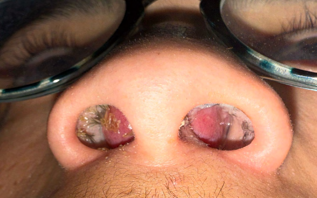
Examination of the nasal pyramid revealed no deformation or visible traumatic lesions. Nasal endoscopy revealed a bilateral, fluctuant swelling of the nasal septum causing complete obstruction of the nasal cavities, with congested nasal mucosa (Figure 1).
Biological tests showed leukocytosis of 14,320/mm³ with neutrophil predominance (8,306/mm³).
An exploratory puncture was performed, yielding 8 cc of pus, followed by incision and drainage under local anesthesia. Intraoperative exploration confirmed the integrity of the nasal cartilage.
Empirical intravenous antibiotic therapy was initiated (amoxicillin-clavulanate), and later tailored to Streptococcus pneumoniae based on culture results. The patient’s clinical condition improved rapidly, and he was discharged after seven days, completing a 14-day antibiotic course.
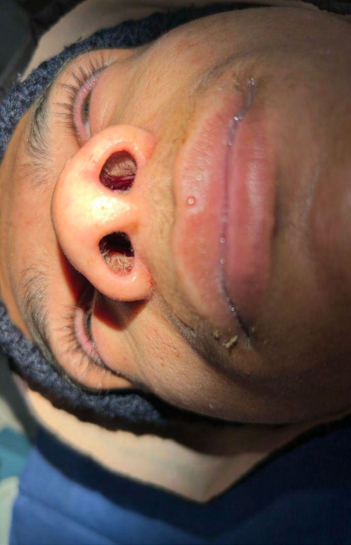
The outcome was favorable, with no recurrence of the abscess. At follow-up, the nasal septum was intact and straight, with good nasal airflow (Figure 2).
Discussion
A nasal septal abscess is defined as a purulent collection located between the septal cartilage or bone and the mucoperichondrium or mucoperiosteum. It is an uncommon condition, with a limited number of cases reported in the literature. However, this rarity appears to be relative, as mild forms often go unnoticed clinically. The condition can occur at any age but is more frequently seen in children, with a higher prevalence among boys [1, 2].
The abscess often results from the superinfection of a septal hematoma, usually following nasal trauma [3]. This trauma may be minor or even unnoticed by the patient, and the interval between the initial event and the development of the abscess typically ranges from five to seven days [3]. Another frequent cause is iatrogenic, secondary to nasal septum surgery [1, 2, 3].
The pathophysiological mechanism involves the rupture of small blood vessels in the septum following facial trauma, leading to a hematoma. This hematoma compromises the vascular supply to the cartilage by separating it from its perichondrium, thereby causing ischemia followed by necrosis. Secondary infection of the hematoma worsens the condition through the action of proteolytic enzymes released by bacteria [1, 2, 4].
Non-traumatic etiologies are rare and mainly concern immunocompromised patients. They are often linked to the infectious spread from the sinuses, particularly the ethmoid air cells or the sphenoidal sinus [1, 5, 6].
The dissemination occurs through the direct diffusion of pus beneath the periosteum, crossing the perpendicular plate of the ethmoid to reach the subperichondral space [1, 2, 5]. More exceptional causes have also been described, such as the spread of a dental or local skin infection (like a nasal furuncle), or a distant infectious focus in immunocompromised patients [1].
Clinical evaluation is a crucial step in diagnosis. It must be performed gently, especially in children, for whom examination under general anesthesia may sometimes be necessary. The combination of anterior rhinoscopy and nasal endoscopy generally reveals a fluctuant bulging of the septal mucosa with an inflammatory appearance, showing a reddish to purplish, or even grayish, discoloration.
This swelling is most often located in the anterior portion of the nasal septum and typically presents bilaterally, although unilateral or posterior locations are also possible. The presence of a fistula draining pus is rare, which can make the diagnosis more challenging.
Clinical examination also aims to identify signs that may indicate the cause, such as a recent nasal trauma or an infectious focus of cutaneous or dental origin. It is also useful for distinguishing an abscess from a simple septal hematoma. The decisive diagnostic procedure remains the aspiration of the fluctuant area, which allows for the extraction of pus, thereby confirming the purulent nature of the lesion and providing a sample for microbiological analysis [4].
Among the most frequently isolated pathogens, Staphylococcus aureus ranks first. Other bacteria such as Streptococcus pneumoniae, Haemophilus influenzae, Streptococcus milleri, Streptococcus viridans, Staphylococcus epidermidis, and anaerobic organisms can also be found. Polymicrobial infections, though less common, are often encountered in immunocompromised individuals [4, 7, 8, 9, 10].
While not essential for making the diagnosis, additional examinations can help determine the etiology. A nasal bone X-ray may reveal a fracture, while a Blondeau view can highlight sinusitis. Panoramic dental radiography is useful for identifying a buccodental infectious origin. Computed tomography (CT) imaging is helpful in assessing the extent of the abscess, the condition of the cartilage and bone, detecting any associated sinusitis, or identifying potential intracranial or vascular complications. Laboratory tests contribute to estimating the severity of the infection and investigating a possible immune deficiency [8, 10].
Therapeutic management primarily relies on effective surgical drainage of the abscess, involving the evacuation of pus and excision of necrotic tissue. This procedure is generally performed under local anesthesia, but general anesthesia is preferred in children for greater comfort and safety. The intervention is followed by bilateral nasal packing, maintained for 48 to 72 hours. Antibiotic treatment is essential, initiated empirically and then adjusted based on the antibiotic susceptibility testing. At the same time, it is crucial to treat the underlying cause as well as any potential complications [1, 3, 4].
A delay in treatment or inadequate management can lead to aesthetic and functional sequelae such as nasal dorsum deformity, septal perforation, or columellar retraction. In children, it can impair the harmonious development of the nasal pyramid. More serious life-threatening complications may also occur, such as intracranial abscess, meningitis, or cavernous sinus thrombophlebitis [3, 5, 6].
Conclusions
Nasal septal abscess is a rare but potentially serious condition that requires prompt diagnosis and intervention to prevent both aesthetic and functional sequelae. This case highlights the importance of considering this diagnosis in the context of recent nasal trauma, even when minor or unnoticed, particularly in patients at risk such as those with neurological disorders. Early drainage combined with appropriate antibiotic therapy allows for rapid resolution and preservation of the nasal structures. Awareness and timely management are essential to avoid complications, especially in pediatric and adolescent patients where long- term nasal development may be compromised.
References
-
Khedim A, Ben Slimene S, Faidi A, Mansour S, Yahia SBH et al. (2007) A case of Nasal septum abscess. Medicine and Infectious Diseases 37(3): S260-S263.
-
Nadour K, Hemmaoui B, Errami N (2010) Nasal Septum Abscess: The ENT and Head and Neck Surgery Letter.
-
Alshaikh N, Stephen LO (2011) Nasal septal abscess in children: from diagnosis to management and prevention. International Journal of Pediatric Otorhinolaryngology 75(6): 737-744.
-
Kryger H, Dommerby H (1987) Haematoma and abscess of the nasal septum. Clinical Otolaryngology & Allied Sciences 122(12): 125-129.
-
Tien DA, Krakovitz P, Anne S (2016) Nasal septal abscess in association with pediatric acute rhinosinusitis. International Journal of Pediatric Otorhinolaryngology 91: 27-29.
-
Hassani R, Aderdour L, Maliki O, Boumed A, Elfakiri MM et al. (2010) Nasal septal abscess complicating acute sinusitisin a child. Archives of Pediatrics 18(1): 15-17.
-
Makitie A, Aaltonen LM, Hytonen, Malmberg H (2000) Postoperative infection following nasal septoplasty. Acta Otolaryngol 543: 165-166.
-
Santiago R, Villalonga P, Maggioni A (1999) Nasal septal abscess: a case report. Inter Pediatr 14: 229-231.
-
Hariri MA, Duncan PW (1989) Infection complications of brief nasotracheal intubation. J Laryngol Otol 103(12): 1217-1218.
-
Collins MP (1985) Abscess of the nasal septum complicating isolated acute sphenoiditis. J Laryngol Otol 99(7): 715-719.
- 4th Branchial Cleft Sinus Anomaly Presenting as Recurrent Thyroid Abscess in A Child: A Case Report
- Parotid Duct Injury Repaired Using an Angiocatheter Stent: A Case Report
- Organization and Functionality of the Referral and Counter-Referral System for ENT Disorders in District Hospitals of N’Djamena, Chad: A Cross-Sectional Analytical Study
- Facial Metastases from a Gastrointestinal Stromal Tumor: A Case Report
- Panorama of Ent Cancers and Literature Review: Epidemiological Profile and Therapeutic Management
- Could Antimicrobial Resistance Prove to Be Both a Threat and an Opportunity for us?