The Occurrence and Diversity of Intestinal Digenetic Trematodes in Stray Cats, Felis Catus, In Egypt
Stray cats act as good indicators of fish-borne trematodes in the environment. Large populations of stray cats, Felis catus, are widespread in Giza, Egypt. Therefore, a total of 47 stray cats from some urban districts in Giza, Egypt were necropsied during the period from December 2018 to April 2019 and their intestines were examined for the detection of digenetic trematodes. Six cats were parasitized with 7 species of trematodes with an overall prevalence of 12.77%. Recovered species were belonging to heterophyids (Heterophyes heterophyes, Pygidiopsis summa, Procerovum varium, Ascocotyle sp.), cyathocotylids (Prohemistomum vivax and Mesostephanus appendiculatus), and echinostomatid (Echinochasmus liliputanus) in prevalences of 6.38% (3/47), 2.13% (1/47), and 6.38% (3/47), respectively. Mixed infections with heterophyids were noticed in two infected cats. Meanwhile, a mixed infection with H. heterophyes and the echinostomatid was recorded in one cat. Moreover, all trematode-infected cats had concomitant infections with cestode and/or nematode parasites. Therefore, veterinarians and authorized agencies must consider great hygienic measures and sanitary control programs to avoid the potential risk and zoonotic importance of such parasites.
Introduction
Stray cats, Felis catus, are strong predators having the ability to consume a variety of prey hosts including mammals, aves and reptiles. Thus, they act as potential reservoir hosts of a wide range of helminth parasites, particularly digenetic trematodes, reflecting both veterinary and medical importance [1, 2, 3]. Populations of stray cats have a random distribution and they live freely in both urban and rural districts, therefore, with a higher opportunity to disseminate infective eggs to the environment [4, 5, 6, 7, 8, 9].
Investigations on digeneans-infected cats have been done globally [10, 11, 12, 13, 14, 15, 16, 17, 18, 19]. Moreover, several species of echinostomatids and heterophyids, including Heterophyopsis continua, Pygidiopsis summa and Heterophyes nocens Echinostoma revolutum, Echinostoma hortense, Echinochasmus japonicus were recorded in stray cats in the Korean Peninsula [2, 20, 21, 22]. In Arabian countries, Schuster, et al. [23] detected Heterophyes heterophyes and Heterophyopsis continua in Dubai, United Arab Emirates. In Kuwait, for the first time, El- Azazy, et al. [24] revealed that 24.6% of cats infected with 14 species of trematodes, predominantly H. heterophyes and H. dispar, Heterophyes nocens Haplorchis taichui, Echinochasmus japonicus, Mesostephanus appendiculatus, Haplorchis yokogawai, and Pygidiopsis genata.
In Egypt, few studies on digenean trematodes infecting cats have been performed previously. Among those, Arafa, et al. [25] reported Heterophyes heterophyes, Echinochasmus perfoliatus and Euparyphium melis in Giza and Cairo provinces. Other species of echinostomatids and heterophyids were recorded in Egyptian literature during the period from 1981 to 2011 [18, 26, 27, 28].
The current study aimed to determine the prevalence of intestinal digenean trematodes recovered from stray cats, Felis catus, necropsied in randomly selected urban districts in Giza province, Egypt with morphological description of the revealed flukes.
Materials and Methods
Sampling Area and Animals
Forty seven stray cats, of both sexes, were obtained from randomly selected urban areas of Giza province (coordinates: 29° 15′ 36″ N, 29° 40′ 12″ E), Egypt during the period from December 2018 to April 2019. Based on dentition, cats aged either less than 3 years or more than 3 years.
In the current study, seven digenetic trematode species were obtained. The recovered trematodes belonged to heterophyids (Heterophyes heterophyes, Pygidiopsis summa, Procerovum varium, Ascocotyle sp.), cyathocotylids (Prohemistomum vivax and Mesostephanus appendiculatus), and echinostomatids (Echinochasmus liliputanus). It has been found that six (12.77%) cats were parasitized; 3 cats were infected with heterophyids, one cat was infected with cyathocotylids and 3 cats were parasitized with the revealed echinostomatid, Echinochasmus liliputanus (Table 1). Among infected cats, 2 individuals (one male and one female) aged 1-3 years and 4 ones (one male and 3 females) aged more than 3 years. Moreover, 4 (females) out of 6 infected cats co- infected with adult tapeworms.
Necropsy of Cats
During necropsy, the abdominal cavity was incised and the entire intestinal tracts of all carcasses were cut, tightly ligated with fine gauze. The small intestine, in particular, was longitudinally opened with a fine scissor and washed in 0.85% saline until the supernatant was cleared. The mucosa was scraped with a scalpel. The epithelial scrapings were passed through 60-80 mesh wire sieves. The contents of the sieves were washed with tap water and sediments were allowed to settle down and carefully examined under a stereomicroscope [3, 29, 30].
Laboratory Procedure
The recovered trematodes were compressed between two slides/cover slips. The flukes were then fixed in 10% neutral buffered formalin, washed with distilled water, and stained with potassium alum carmine (5 min for small- sized helminths and up to 4 hours for large helminths). Dehydration occurs in ascending grades of ethyl alcohol (70,80,90,95, and 100%). Then, specimens were cleared in xylene, and mounted with Canada balsam [30, 31].
Identification of Trematodes
Prepared slides were carefully examined under a light microscopy using different magnifications, and recovered helminths were identified according to keys given by Yamaguti, [32] and Soulsby EJL [29]. Available photographs were taken using a digital microscope (Leica microsystems, CH-9435 Heebrugg, Ec3, and Singapore). Measurements of the recovered digenean trematodes were in micrometers.
Results
| Digenetic trematode | Infected cats (n= 47) | Prevalence | Worm burden |
|---|---|---|---|
| Heterophyes heterophyes | 3*§ | 6.38 | 1-10 |
| Pygidiopsis summa | 1* | 2.13 | One |
| Procerovum varium | 1* | 2.13 | One |
| Ascocotyle longicollis | 1* | 2.13 | 2 |
| Prohemistomum vivax | 1** | 2.13 | 2 |
| Mesostephanus appendiculatus | 1** | 2.13 | 1-12 |
| Echinochasmus liliputanus | 3§ | 6.38 | 1-5 |
Table 1: ** The prevalence and worm burden of digenetic trematodes recovered from the intestinal tracts of stray cats in Giza, Eg
*One cat had mixed infections of heterophyids. One cat had a mixed infection of both P. vivax and M. appendiculatus. §One cat had a mixed infection of H. heterophyes and E. liliputanus Table 1:** The prevalence and worm burden of digenetic trematodes recovered from the intestinal tracts of stray cats in Giza, Egypt.
Species: Heterophyes heterophyes Diagnosis (Figures 1 a,b)
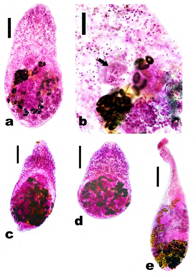
Figure 1: Adult heterophids recovered from necropsied cats. a Adult Heterophyes heterophyes. Scale bar = 100 μm. b Gonotyle (ventrogenital complex) (arrow) of adult Heterophyes heterophyes showing numerous rodlets (more than 70). Scale bar = 50 μm. c Adult Pygidiopsis summa. Scale bar = 100 μm. d Adult Procerovum varium. Scale bar = 100 μm. e Adult Ascocotyle longicollis. Scale bar = 100 μm.
Short pear-shaped fluke measured 0.8-1.1 x 0.25 mm. It was wider posteriorly. The oral sucker measured approximately 50 μm in diameter. The oesophagus measured 0.2 mm long. The ventral sucker is situated immediately anterior to the middle. The genital sucker lies directly behind it and to one side and bears an incomplete circle of 70-80 small rods. The testes are oval and horizontal in position measured about 50 μm.
Species: Pygidiopsis summa Diagnosis (Figure 1c)
Very small fluke measured 0.45 x 0.20 mm. The body is more or less pointed at the fore end and broad at the hind end. Adult are ventrally concave, pyriform-shaped with oral and ventral suckers as well as a genital apparatus. The oral sucker measured about 37.5 μm. The oesophagus measured 57.0 μm. The whole body surface is covered with numerous tegumental spines, and sensory papillae.
Species: Procerovum varium Diagnosis (Figure 1d) The body is small pear-shaped measured 0.35 × 0.20 mm with the greatest width at the posterior third of the body. The cuticle is provided with fine spines particularly at the anterior half. The oral sucker is subterminal and measured 50.0 μm. The prepharynx is very short. Pharynx is subglobular or elliptical-shaped. The oesophagus is short and measured 0.12 mm long. The ventral sucker is very small, located just behind the bifurcation of intestinal ceca, embedded in the ventrogenital sac and masked by several eggs. Intestinal ceca bifurcate at the level of anterior third and terminate at the posterior fourth of the fluke. The ovary is spherical/ovoid, measured 50.0 μm and located sagital to the ventral sucker. Two testes, each is subglobular-shaped, located at the middle of the hind body and measured 95.0 × 99.0 μm. Vitellaria are greatly follicular and located in the posterior testicular field at the posterior extremity. The uterus is filled with small yellowish eggs (measured 22.0 × 11.0 μm) and occupies the entire area of the hind body.
Species: Ascocotyle (Phagicola) longicollis Diagnosis (Figure 1e) A small elongate conical-shaped fluke, measured approximately 0.60 x 0.18 mm. The oral sucker measured 37.5 x 50.0 μm and bears approximately 18-20 hooklets arranged in 2 rows. The pharynx measured 25.0 x 27.5 μm and leads into the conical prepharynx that is characteristic of this genus. The oesophagus measured 0.18 mm. The genital opening is just anterior to the ventral sucker which measured 30.0 x 37.5 μm. The majority of the reproductive organs are located posterior to the ventral sucker. Paired testes are symmetrical and located at the posterior end of the body measuring 50.0 x 62.5 μm. The egg is oval-shaped and measured 12.5 x 25.0 μm.
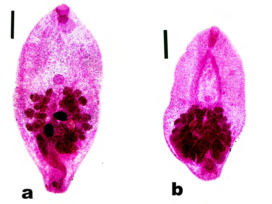
Species: Prohemistomum vivax Diagnosis (Figure 2a) The adult fluke measured 1.2 mm x 0.55 mm. The body is oval rather than elongate. The cuticle is covered with minute spines which gradually diminish in size towards the posterior end. A sub terminal and rounded oral sucker measured 70.0 x 70.0 μm. The ventral sucker (50 x 50 μm) is weakly developed, oval-shaped and lies at the junction of the anterior third with the middle third of the body. Testes are ovoid-shaped and smooth and they lie posteriorly, obliquely tandem at the posterior third of the body. Ovoid ovary and lies between the two testes. Vitelline follicles are voluminous and filling the posterior half and arranged in an interrupted horse-shoe- shaped manner around the gonads and the upper half of the cirrus pouch. At the posterior end, there is a distinct cirrus pouch (0.1 x 0.35 mm) along with a narrow tubular vagina parallel to it.
Species: Mesostephanus appendiculatus Diagnosis (Figure 2b) Linguiform-shaped fluke, measured 1.12 × 0.35 mm. The anterior part is blunt and the posterior part is cylindrical, wider and provided with protruded caudal appendage (measured 0.12 mm long). The oral sucker is subterminal and measured 50 μm. The pharynx is somewhat large, more or less close to the oral sucker and measured 50 × 40 μm. The oesophagus is partially covered by the pharynx and measured 0.10 mm. The intestinal bifurcation begins at the anterior fourth and the posterior part of them is completely hidden by vitelline glands. Testes are oval/ovoid, tandem at the posterior fourth of the body and measured 80 μm in diameter. Few large eggs which are yellowish to brown and located at the posterior part. The ovary measured 60 μm in diameter and located in between the 2 testes and masked by vitelline glands.
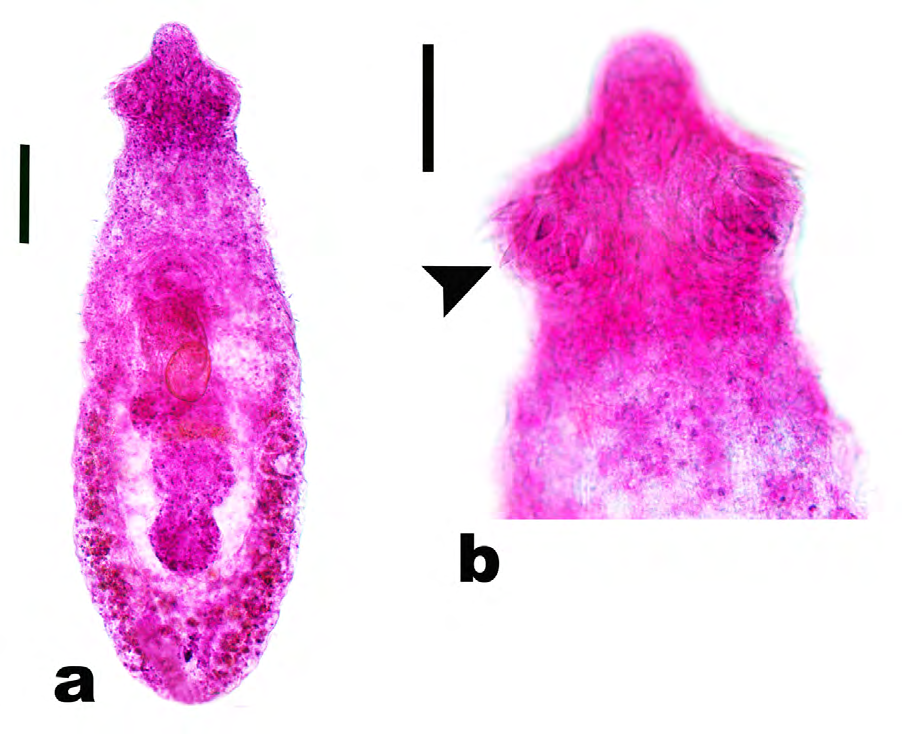
Species: Echinochasmus liliputanus Diagnosis (Figure 3) A small, elongate and leaf-shaped fluke, with its extremities narrower than the middle of the body. Sharply pointed spines are confined to the anterior part of the cuticle. The length of the body measured 0.50-0.70 mm (average 0.60 mm). The maximum width (0.20 mm) was at the level of the testes. The head collar measured 112.5 x 50 μm and armed with 24 unequally-sized collar spines arranged in a single dorsally interrupted row (each spine measured approximately 25.0 μm). The oral sucker is rounded-shaped, subterminal and measured 40.0 μm. The ventral sucker is approximately twice the size of the oral sucker measuring 75.0 μm in diameter. The oesophagus is short measuring 100 μm long. The two testes are tandem closely related in posterior half of the body measuring 60.0 μm wide. The fore testis is more or less larger than the hind one. The ovary is oval-shaped, anterior to the testes and measured 70.0 x 50.0 μm. Vitellaria are large follicles extended from the middle to the posterior extremity. Eggs are few (2-6), golden brown with a distinct knob and measured 30.0 x 14.0 μm.
Discussion
Felids, particularly cats, may harbor several species of digenetic trematodes. The majority of them are zoonotic with subsequent potential harms to humans, especially in low-income countries [25].
In Egypt, helminths, particularly trematodes, of stray cats are considerably existed due to the higher opportunities of the heterogenic life cycle of those trematodes, as cats (the final hosts) have a predatory behavior that permit other reptiles, mammals and birds, containing the infective stages, to be ingested [3, 16, 23, 33].
Currently, the prevalence of digenetic trematodes was 12.77% (6/47) in Giza province. In Egypt, previous literature reported higher infection rates; Fouly EA, [27] found that the prevalence of flukes-induced infection was 42.0%, Thabit HTM, [28] who reported that the prevalence was 61.6% and El-Dakhly, et al. [3] who revealed that the prevalence was 12.9%. Similarly, in Kuwait, El-Azazy, et al. [24] recorded a prevalence of 24.6%. Authors suggest that the lower prevalence in the current study might be referred to the enhancing hygienic measures and strict governmental rules that permit the hygienic disposal of garbage, containing various remnants of infected fish, as well as the periodical elimination of stray cats, the definitive and intermediate hosts of digenetic trematodes.
The present work revealed 7 digenetic trematodes. In Egypt, Kuntz RE, et al. [34] recorded 14 digenetic trematodes, Heterophyes heterophyes, Heterophyes aequalis, H. pumilio, H.
taichui, H. yokogawai, S. falcatus, Pygidiopsis genata, Phagicola longicollis, Phagicola ascolonga, Stictodora sawakinensis, Echinochasmus liliputanus, Stephanoprora denticulatoides, Mesostephanus appendiculatus, and Cynodiplostomum namrui, from domestic cats. They added that the presence of these flukes might be due to ingesting fishes infected with metacercariae. Therefore, stray cats in those areas are potential definitive hosts for various digenetic trematodes.
Heterophyids are often intestinal flukes of fish-eating mammals and their metacercariae encyst in tissues of fishes [35, 36, 37]. Herein, the most frequent digenetic trematodes belonged to the family Heterophyidae. Coinciding with such finding, El-Azazy, et al. [24] recorded H. heterophyes and H. dispar, suggesting that stray cats are good biomarkers of fish-borne digenetic trematodes. Moreover, Eom, et al. [20] revealed Heterophyes continua, H. nocens and Pygidiopsis summa from cats purchased at Jungang Market in Seoul, South Korea. Cort WW, et al. [38] mentioned the morphological characters of Egyptian heterophyids. The number of chitinous rodlets in the genital sucker (gonotyl) was 75- 85 in Heterophyes heterophyes and 50-60 in H. heterophyes nocens, a characteristic feature for the two heterophyids. The close association between snails and brackish water fish, intermediate hosts for such heterophyids, explain the occurrence of those flukes. In terms of zoonosis, Chai, et al. [39] stated that heterophyids might induce erratic parasitism in humans, and their eggs could be found within inflammatory reactions of affected organs with further complications resulted from the release of eggs into the blood stream via the intestinal wall.
Among heterophyids, Ascocotyle (syn. Phagicola) species have unique morphological character; the anterior end of adults has a crown of double rows of spines (14-17) and a solid prolongation of the oral sucker [40]. A clear association between the existence of A. longicollis, fish and predators was observed [41]. Pygidiopsis summa is a heterophyid fluke having a rapid development inside the final hosts [39]. In the present investigation, the occurrence of P. summa indicated the existence of definitive hosts, stray cats, and intermediate hosts, fish.
Among cyathocotylids, Mesostephanus appendiculatus is a common fluke of major public health significance, thus, it is considered a zoonotic fish-borne digenetic trematode [42]. Such fluke requires more than one host; fish eating mammals/birds (definitive host), gastropods and fish (intermediate hosts) [43]. The feeding habits of stray cats as free living animals evidently suggest the occurrence of M. appendiculatus. Meanwhile, Prohemistomum vivax, is a cyathocotylid needs catfish (a predominant consumed fish in Egypt) as intermediate host [44]. Currently, adult Prohemistomum vivax was revealed from stray cats.
Echinochasmus liliputanus is an intestinal echinostmatid in canids, felids, and humans. Herein, it was detected in 6.45% of infected cats. On the other hand, Thabit, [28] and Fang, et al. [9] revealed a higher prevalence (28.3%). Drinking un boiled pond water infected with cercariae is the often route of human infection [45, 46].
To minimize the risk of potential and zoonotic trematode infections, veterinarians, parasitologists and biologists are advised to carefully consider the importance of snails, reptiles and fish as eminent intermediate hosts for digenetic trematodes as well as the hygienic disposal of those hosts to reduce the possibility of completing life cycles of the flukes. Meanwhile, authoritive agencies are strongly recommended to eliminate strays cats and dogs.
Conflict of Interest
Authors declare that there is no conflict of interest.
Ethical Standards
Animals used in the present study were obtained as a result of the cooperation between the Directorate of Veterinary Medicine, Beni-Suef, Egypt and the Faculty of Veterinary Medicine, Beni-Suef. The current work was done according the ethics Animal and Human Research Committee of the Faculty of Veterinary Medicine, Beni-Suef University, Egypt.
References
-
Millan J, Casanova J (2009) High prevalence of helminth parasites in feral cats in Majorca island (Spain). Parasitol Res 106: 183-188.
-
Chai JY, Bahk YY, Sohn WM (2013) Trematodes recovered in the small intestine of stray cats in the Republic of Korea. Korean J Parasitol 51(1): 99-106.
-
El-Dakhly Kh M, Aboshinaf ASM, El-Nahass E, Gharib AF (2017) Preliminary study on the helminth fauna in necropsied stray cats (Felis catus) in Beni-Suef, Egypt. J Adv Vet Res 7(4): 87-92.
-
Shahram J, Meshki B, Meshki M (2002) A study of helminthic infection of gastrointestinal tract in stray cats at urban areas in Isfahan. J Vet Res 57(2): 25-27.
-
Bahadori Sh R, Eslami A, Meshgi B, Poor Hosseini S (2004) Study on stray cats infected with parasitic helminthes in Tehran. J Fac Vet Med Univ Tehran 59: 171-174.
-
Sharif M, Nasrolahei M, Ziapour SP, Gholami S, Ziaei H, et al. (2007) Toxocara cati infections in stray cats in northern Iran. J Helminthol 81(1): 63-66.
-
Zibaei M, Sadijadi SM, Sarkari B (2007) Prevalence of Toxocara cati and other intestinal helminthes in stray cats in Shiraz, Iran. Trop Biomed 4(2): 39-43.
-
Mohsen A, Hossein H (2009) Gastrointestinal parasites of stray cats in Kashan, Iran. Trop Biomed 26(1): 16-22.
-
Fang F, Li J, Huang T, Guillot J, Huang W (2015) Zoonotic helminths parasites in the digestive tract of feral dogs and cats in Guangxi, China. BMC Vet Res 11: 211.
-
Gregory GG, Munday BL (1976) Internal parasites of feral cats from the Tasmanian Midlands and King Island. Aust Vet J 52(7): 317-320.
-
Min HK (1981) An epidemiological study on zoonoses in Korea. Korean J Parasitol 19(1): 60-75.
-
Huh S, Sohn WM, Chai JY (1993) Intestinal parasites of cats purchased in Seoul. Korean J Parasitol 31(4): 371- 373.
-
Scholz T, Uhlírová M, Ditrich O (2003) Helminth parasites of cats from the Vientiane province, Laos, as indicators of the occurrence of causative agents of human parasitoses. Parasite 10(4): 343-350.
-
Labarthe N, Serrao ML, Ferreira AM, Almeida NK, Guerrero J (2004) A survey of gastrointestinal helminth in cats of the metropolitan region of Rio de Janeiro, Brazil. Vet Parasitol 123(1-2): 133-139.
-
Adams PJ, Elliot AD, Algar D, Brazell RI (2008) Gastrointestinal parasites of feral cats from Christmas Island. Aust Vet J 86(1-2): 60-63.
-
Millan J, Casanova J (2009) High prevalence of helminth parasites in feral cats in Majorca island (Spain). Parasitol Res 106(1): 183-188.
-
Borthakur SK, Mukharjee SN (2011) Gastrointestinal helminths in stray cats (Felis catus) from Aizawl, Mizorom, India. Southeast Asian J Trop Med Public Health 42(2): 255-258.
-
Khalafalla RE (2011) A survey study on gastrointestinal parasites of stray cats in northern region of Nile Delta, Egypt. PloS One 6(7): e20283.
-
Zain SN, Shimin N, Pal P, Lewis JW (2013) Macroparasite communities in stray cat populations from urban cities in Peninsular Malaysia. Vet Parasitol 196(3-4): 469-477.
-
Eom KS, Son SY, Lee JS, Rim HJ (1985) Heterophyid trematodes (Hetrophyis continua, Pygidiopsis summa and Heterophyes heterophyes nocens) from domestic cats in Korea. Korean J Parasitol 23(2): 197-202.
-
Sohn WM, Chai JY (2005) Infection status with helminthes in feral cats purchased from a market in Busan, Republic of Korea. Korean J Parasitol 43(3): 93-100.
-
Shin SS, Oh DS, Ahn KS, Cho SH, Lee WJ, et al. (2015) Zoonotic intestinal trematodes in stray cats (Felis catus) from Riverside areas of the Republic of Korea. Korean J Parasitol 53(2): 209-213.
-
Schuster RK, Thomas K, Sivakumar S, O’Donovan D (2009) The parasite fauna of stray domestic cats (Felis catus) in Dubai, United Arab Emirates. Parasitol Res 105(1): 125-134.
-
El-Azazy OM, Abdou NM, Khalil AL, Al-Batel MK, Majeed QA, et al. (2015) Potential zoonotic trematodes recovered in stray cats from Kuwait municipality, Kuwait. Korean J Parasitol 53(3): 279-287.
-
Arafa MS, Nasr NT, Khalifa R, Mahdi AH, Mahmoud WS, et al. (1978) Cats as reservoir hosts of Toxocara and other parasites potentially transmissible to man in Egypt. Acta Parasitol Pol 25: 383-389.
-
Fahmy MA, Khalifa R, Sakla AA (1981) Study on two Echinochasmid parasites (Trematoda: Echinochasmidae) from upper Egyptian cats. Assiut Vet Med J 8: 73-75.
-
Fouly EA (1997) Studies on parasites of cats in Assiut Governorate. Ph.D. Thesis Fac. Vet Med, Assiut Univ, Egypt.
-
Thabit HTM (2011) Biological and parasitological studies on some endoparasites of cats in Assiut, Egypt. PhD. Thesis, Faculty of Veterinary Medicine Assiut University, Egypt.
-
Soulsby EJL (1982) Helminthes, Arthropods and Protozoa of domesticated animals. 7th (Edn.), London, Bailliere, Tindall and Cassel.
-
El-Dakhly Kh M, El-Nahass E, Uni S, Tuji H, Sakai H, et al. (2012) Levels of infection of gastric nematodes in a flock of great cormorants (Phalacrocorax carbo) from Lake Biwa, Japan. J Helminthol 86(1): 54-63.
-
Rozsa L, Reiczigel J, Majoros G (2000) Quantifying parasites in samples of hosts. J Parasitol 86(2): 228-232.
-
Yamaguti S (1958) Systema Helminthum Volume I. The Digenetic Trematodes of vertibrates-part- II. New York, Interscience Publishers Inc.
-
Shaw J, Dunsmore J, Jakob-Hoff R (1983) Prevalence of some gastrointestinal parasites in cats in the Perth area. Aust Vet J 60(5): 151-152.
-
Kuntz RE, Chandler AC (1956) Studies on Egyptian trematodes with special reference to the heterophyids of mammals. I. Adult flukes, with descriptions of Phagicola longicollis sp., Cynodiplostomum namruin. sp., and a Stephanoprora from cats. J Parasitol 42: 445-459.
-
Sepulveda MS, Spalding MG, Kinsella JM, Forrester DJ (1996) Parasitic helminths of the little blue heron, Egretta caerulea, in southern Florida. J Helminthol Soc Wash 63(1): 136-140.
-
Sepulveda MS, Spalding MG, Kinsella JM, Forrester DJ (1999) Parasites of the great egret (Ardea albus) and a review of the helminths reported for the species. J Helminthol Soc Wash 66(1): 7-13.
-
Scholz T, Aguirre-Macedo ML, Salgado-Maldoonado G (2001) Trematodes of the family Heterophyidae (Digenea) in Mexico: a review of species and new host and geographical records. J Nat Hist 35(12): 1733-1772.
-
Cort WW, Yokogawa S (1921) A new human trematode from Japan. J Parasitol 8(2): 66-69.
-
Chai JY, Seo BS, Lee SH, Hong ST (1986) Growth and development of Pygidiopsis summa in rats and mice with a supplementary note on its morphological characters. Korean J Parasitol 24(1): 55-62.
-
Nunez MO (1993) Life-history studies of heterophyid trematodes in the Neotropical Region: Ascocotyle (Phagicola) diminuta (Stunkard and Haviland, 1924) and A. (P.) angrense Travassos, 1916. Syst Parasitol 24: 191-199.
-
Hutton RF, Sogandares-Bernal F (1958) Variation in the number of oral spines of Phagicola longicollis. J Parasitol 44(6): 627-632.
-
El-Bahy NM, Bazh EK, Sorour SS, Elhawary NM (2017) Molecular characterization of the unique Mesostephanus appendiculatus (Trematoda: Cyathocotylidae) by small ribosomal RNA from Egypt. Parasitol Res 116(4): 1129- 1136.
-
Gibson DI, Jones A, Bray RA (2001) Keys to the Trematoda, Volume 1. CABI Publishing and the Natural UK, pp: 201.
-
Alghabban AJ (2014) Fish Farms as a source for parasites transport: Parasitological and developmental studies of Prohemistomum vivax with the ameliorating role of Moringa oleifera in the treatment. J Am Sci 10(4): 6-14.
-
Xiao X, Lu DB, Wang TP, Gao JF, Wu WD (1994) Epidemiological studies on Echinochasmus liliputanus infection I. Parasite infection and distribution in final hosts. Chinese J Parasit Dis 7: 285-287.
-
Sohn WM, Eom KS, Min DY, Rim HJ, Hoang EH, et al. (2009) Fish borne trematode metacercariae in freshwater fish from Guangxi Zhuang Autonomous Region, China. Korean J Parasitol 47(3): 249-257.
- California Red-Legged Frog and Non-Listed Amphibians Response to Non-Native Fish Removal
- Industrial Standardization of the Bio-OS: Algorithmic Codification of Resilience Engineering Guidelines and Version V8 Architecture
- Climate Variability and the Sustainability of Snail Farming in Nigeria: Past Trends, Present Challenges and Potential Outlook
- The Evaluation of the Surveillance System of Anthrax in Gilgit-Baltistan, Pakistan, 2018
- Natural Decline to Extinction of A New Zealand Rabbit Population
- Mitochondrial Bio-Logistics: Steering Co-Enzyme Q10 and Lycopene Synergies within the Science 4.0 Bio-OS Framework