Histopathological Studies of Gills, Liver and Gonads of Clarias gariepinus and Oreochromis niloticus Collected from Rivers Ureje and Ogbese in Ado-Ekiti, Nigeria
This study investigated the histopathology of the gills, liver and gonad of Clarias gariepinus and Oreochromis niloticus collected from Rivers Ureje and Ogbese in Ekiti state. The results obtained from the analysis of water sampled from the two aquatic environments indicate that the quality of water varies greatly between the two study areas. Histological alterations were observed in the liver, gills and gonad of fish from both rivers, including circulatory disturbances such as telangiectasia and epithelial lifting, hyperplasia of mucous cells and epithelial cells between secondary lamellae, structural alterations in the form of fusion and branching of primary and secondary lamellae, and regressive changes in the form of intracellular alterations within gill epithelial cells.
Introduction
Aquatic organisms, including fishes, accumulate pollutants directly from contaminated water and indirectly via the food chain. The organ most associated with the detoxification and biotransformation process of toxicants including metals is the liver; because the liver of fish can be considered a target organ to pollutants, alterations in its structure can be significant in the evaluation of fish health and exhibit the effects of a variety of environmental pollutants [1, 2]. In aquatic ecosystem, fish are considered as sentinel organisms for evaluating toxicosis of xenobiotic substances under aquatic environment. Fish occupies the top of the aquatic food chain, are highly visible resource and highly exposed to accumulate these toxic substances [3]. In addition to this, they are in direct contact continuously with these substances in water via their gills and their body surface. The physical and chemical changes in aqueous environment often cause some physiological changes in fish, thus, the water quality of an aquatic body is very crucial because it determines the productivity and other parameters necessary for fish survival. Many countries including Nigeria have legislated against the use of chemical poisons in aquatic systems and instead have policies favoring the use of natural bio–degradable alternatives to remove unwanted fish species in aquatic systems [4, 5]. Histopathological investigations have long been recognized to be reliable biomarkers of stress in [6]. Histopathological changes have been widely used as biomarkers in the evaluation of the health of fish exposed to contaminants, both in the laboratory and field studies. One of the great advantages of using histopathological biomarkers in environmental monitoring is that this category of biomarkers allows examining specific target organs, including gills, liver, and gonads that are responsible for vital functions, such as respiration, excretion, reproduction and the accumulation and biotransformation of xenobiotics in the fish [7].
Furthermore, the alterations found in these organs are normally easier to identify than functional ones [8], and serve as warning signs of damage to animal health [9]. Histological abnormalities in fish exposed to pollutants include impaired growth and reproduction, which reduces the fish population and possible extinction of fish at continuous exposure. Histopathological alterations can be used as indicators for the effects of various anthropogenic pollutants on organisms and are a reflection of the overall health of the entire population in the ecosystem [10]. These histopathological biomarkers are closely related to other biomarkers of stress since many pollutants have to undergo metabolic activation in order to be able to provoke cellular change in the affected organism.
This research therefore investigated the histopathology of the gills, liver and gonads of Clarias gariepinus and Oreochromis niloticus collected from Rivers Ogbese and Ureje which are continuously exposed to domestic and agricultural wastes.
Materials and Methods
Study Area
The study was carried out in Rivers Ogbese and Ureje both of which are located in Ado- Ekiti Local Government Area, Ekiti State. River Ureje is located between Latitudes 07°35’ and 07°40’N and Longitudes 005°10’and 005°15’E while River Ogbese lies between Latitudes 07°30’ and 07°40’N and Longitudes 005°15’ and 007°20’ E.
Collection of Samples
Clarias gariepinus and Oreochromis niloticus were collected by local fishermen using gill nets from the two locations. The fish samples were transported to the laboratory in well aerated containers 25% filled with water. Samples of water were collected from the rivers for physico- chemical parameters and heavy metal analyses.
Preparation of Samples
The abdominal walls of the fish were dissected open through a mid-ventral abdominal incision. The positions of the organs were carefully noted in situ. The individual gills, liver and gonads were carefully dissected. The organs are stored in 10% formalin solution in the specimen containers.
Tissues Processing, Staining and Histopathological Studies
The preserved organs (gills, liver, gonads) were used for histopathological studies. The organs were removed from the formalin solution and rinsed in normal saline solution. Samples of the gills, liver, testes and ovary were later fixed in Bouin’s fluid fixative for 72 hrs. Each of the tissue samples taken from the gills, liver and gonads were immersion- fixed separately in 10% neutral buffered formalin at room temperature. The specimens were left in the fixative for 24 h. Following fixation, the tissues were rinsed in running water, dehydrated by immersing in ascending grades of ethyl alcohol, cleared in two changes of absolute xylene, impregnated with molten paraffin wax in hot oven and embedded in paraffin blocks at room temperature.
The paraffin blocks were sectioned with microtome at 5-6µ thickness. Ribbons containing every 8th to 10th sections were collected and gently floated on a tissue floatation bath at 40°C and put on glass microscopic slides. Before staining, the tissue sections were deparaffinized with xylene and hydrated with ethanol. After rinsing the slides with distilled water, sections were stained regressively with Haematoxylin for 10 min. The sections were washed in tap water and dip into 1% of acid alcohol for differentiation and to remove excess stain. The sections were rinsed briefly in running tap water to remove excess acid and halt distain. The slides were then placed in saturated sodium bicarbonate solution for 3 min, and counterstained in 1% alcoholic eosin for 1 min. The Haematoxylin and Eosin stained sections were dehydrated by increasing concentration of ethanol and cleared in xylene and were mounted using DPX mountant and glass cover slips. Microscopic slides were examined under light microscope. Photomicrographs of selected slides of each of the organs under study were taken using Digital Photo Camera mounted on a binocular compound microscope (Axiostar MWIB, US).
Results
Histopathological Investigations of Clarias gariepinus and Oreochromis niloticus from River Ureje and River Ogbese
The histological sections of the various organs (liver, gills and gonads) of Clarias gariepinus and Oreochromis niloticus from River Ureje and River Ogbese are presented in Plates 1-3.
Sections of the Fish Gills
Plate 1 shows the histological sections of a normal fish gills as well as fish gills of Clarias gariepinus and Oreochromis niloticus from River Ureje and River Ogbese respectively. Plate 1a shows the photomicrograph of the gills of a healthy fish revealing the primary lamellae consisting mainly of epithelial tissue, cartilage and a vascular system. The secondary lamellae protrude along the entire length of the primary lamellae, each consisting of blood arterioles through which oxygen exchange with the surrounding water takes place. Plate 1b shows the photomicrograph of the gills of Clarias gariepinus collected from River Ureje showing normal architecture of gills with intact primary gill lamellae filament (PGL) and secondary gill lamellae (SGL), gill arches and gills rays with no pathological lesions. Plate 1c shows the photomicrography of the gills of Clarias gariepinus collected from River Ogbese showing extensive necrosis in the covering epithelium of the secondary lamella with leukocytes aggregation and showing necrosis and leukocytes infiltration in the epithelium of suprabronchial organs. Plate 1d shows the photomicrograph of the gills of Oreochromis niloticus collected from River Ureje which shows the vasodilation, hyperplasia and partial fusion of lamellae. Plate 1e shows the photomicrograph of the gills of Oreochromis niloticus collected from River Ogbese which shows haemorrhage in primary lamellae and separation and degeneration in epithelial cells of secondary lamellae.
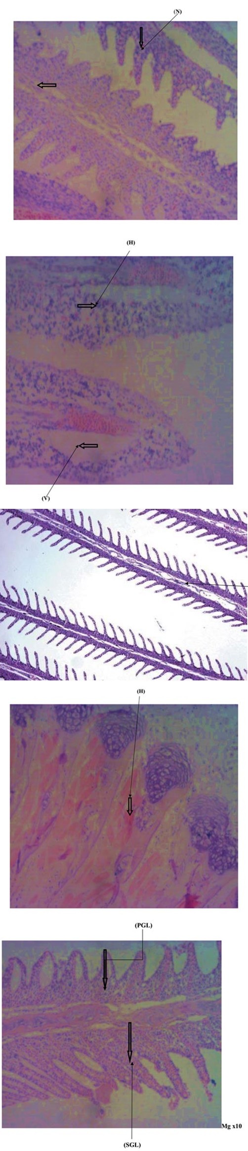
Plate 1a: Photomicrograph of the gills of a healthy fish.
Plate 1b: Photomicrograph of the gills of Clarias gariepinus collected from River Ureje.
Plate 1c: Photomicrograph of the gills of Clarias gariepinus collected from River Ogbese.
Plate 1d: Photomicrograph of the gills of Oreochromis niloticus collected from River Ureje.
Plate 1e: Photomicrograph of the gills of Oreochromis niloticus collected from River Ogbese.
Sections of the fish liver
Plate 2 shows the histological sections of a normal fish liver as well as fish liver of Clarias gariepinus and Oreochromis niloticus from River Ureje and River Ogbese respectively. Plate 2a shows the photomicrograph of the liver of a healthy fish revealing a normal hepatic architecture consisting mainly of highly vascularized epithelial tissue and hepatocytes. Plate 2b shows the photomicrograph of the liver of Clarias gariepinus collected from River Ureje showing a normal structure of liver hepatocytes and nucleus. Plate 2c shows the photomicrography of the liver of Clarias gariepinus collected from River Ogbese showing t h e degradation of cellular hepatocytes, sinusoids with pyknotic nuclei. Plate 2d shows the photomicrograph of the liver of Oreochromis niloticus collected from River Ureje which shows normal liver histology in control fish showing normal hepatocytes and pancreas, loss of contact between hepatocyte and pancreocytes. Plate 2e shows the photomicrograph of the liver of Oreochromis niloticus collected from River Ogbese which shows dilatation and blood congestion. It also shows melano- macrophage aggregation, hepatocyte hypertrophy, nuclear pyknosis and cellular degeneration, intravascular heamolysis in hepaportal blood vessels.
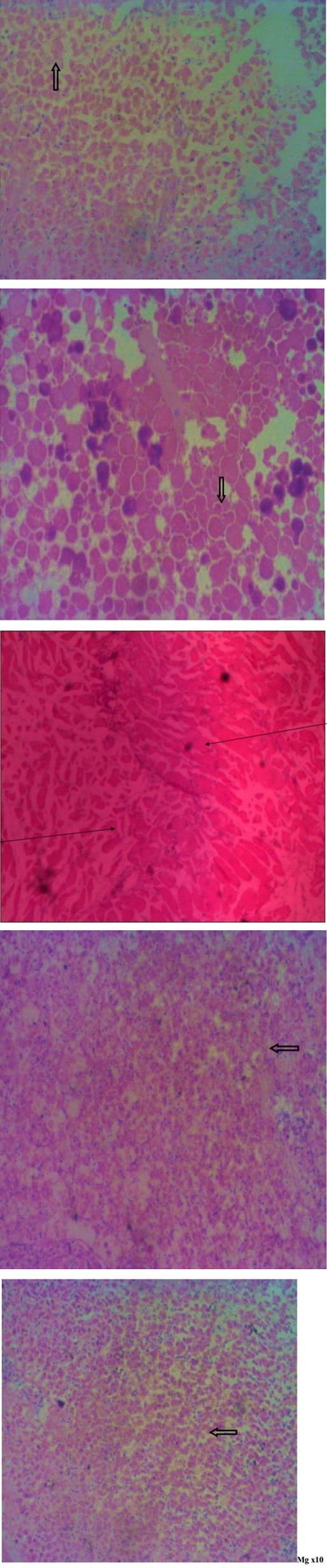
Plate 2a: Photomicrograph of the liver of a healthy fish.
Plate 2b: Photomicrograph of the liver of Clarias gariepinus collected from River Ureje.
Plate 2c: Photomicrograph of the liver of Clarias gariepinus collected from River Ogbese.
Plate 2d: Photomicrograph of the liver of Oreochromis niloticus collected from River Ureje.
Plate 2e: Photomicrograph of the liver of Oreochromis niloticus collected from River Ogbese.
Sections of the fish gonads
Plate 3 shows the histological sections of a normal fish gonad as well as fish gonad of Clarias gariepinus and Oreochromis niloticus from River Ureje and River Ogbese respectively. Plate 3a shows the photomicrograph of the gonad of a healthy fish revealing nuclei of epithelia cells and interstitial connective tissue and the proximal part showing the presence of numerous glandular cells and smooth muscles cells.
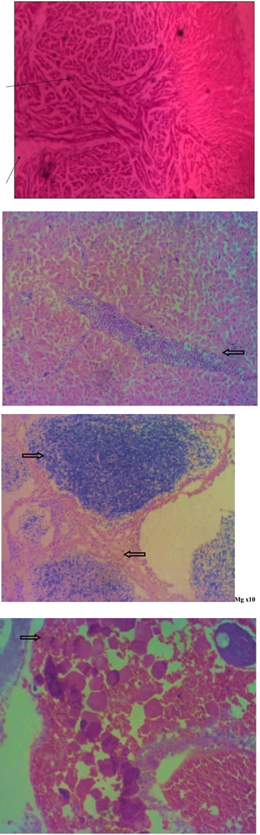
Plate 3a: Photomicrograph of the gonad of a healthy fish.
Plate 3b: Photomicrograph of the gonad of male Clarias gariepinus collected from River Ureje.
Plate 3b shows the photomicrograph of the gonad of male Clarias gariepinus collected from River Ureje showing lobules filled with colloidal substance, nuclei of epithelia cells and interstitial connective tissue and the proximal part showing the presence of numerous glandular cells and smooth muscles cells. Plate 3c shows the photomicrograph of the gonad of female Clarias gariepinus collected from River Ogbese showing tissue with few previtellogenic oocytes dispersed amongst abnormal tissue containing numerous oogonia, including multinucleate stages. Plate 3d shows the photomicrograph of the gonad of female Oreochromis niloticus collected from River Ureje showing many atretic follicles, some extent of damage can be seen in the architecture of tunica albuginea as they become loose, stromal haemorrhage, lymphocytic infiltration and alteration in germinal epithelium. Plate 3e shows the photomicrograph of the gonad of female Oreochromis niloticus collected from River Ogbese showing an increase in the amount of stromal and lymphocytic infiltration.
Plate 3c: Photomicrograph of the gonad of female Clarias gariepinus collected from River Ogbese
Plate 3d: Photomicrograph of the gonad of female Oreochromis niloticus collected from River Ogbese.
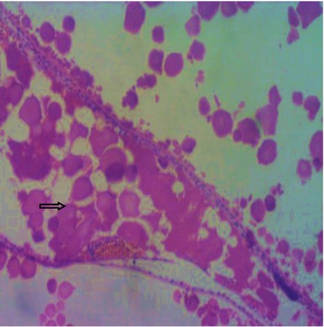
Plate 3e: Photomicrograph of the gonad of female Oreochromis niloticus collected from River Ogbese.
Discussion
Fishes require energy for respiration, reproduction, locomotion, blood chemistry and digestion. There are a number of ways that fish can utilize to make use of oxygen from the water, but the most common means involves the gills. If the water in which they inhabit is polluted with effluents such as agricultural, industrial and domestic wastes, the gills, liver and other vital organs are prone to get damaged. Gills are highly susceptible to toxic chemicals of environmental pollutants, because of direct contact between gills and environmental pollutants .The absorption of toxic chemicals through gills is enhanced by increasing the permeability to water and ions of gill epithelium and by inhibition of ions exchange activity of the chloride cells [11]. The measurements of the effect of aquatic pollutants in freshwater habitats have frequently been tested in gill [12, 13]. Histological study of the gills shows a typical structural organization of the lamella in the untreated fish. However, fish exposed to lead acetate shows several histological alterations, included hypertrophy, lifting of the epithelial linings from the surfaces of secondary lamellae, and at few places, degeneration of lamellar epithelium. Similar observations were reported due to exposure of other freshwater fish to lead and copper [14, 15, 16, 17, 18]. Lifting and hyperplasia of lamellar epithelium could be interpreted as defense responses of the fish, as these alterations increase the distance across which waterborne irritants must diffuse to reach the blood stream [19]. The most common cause of cellular degeneration in gill filaments is oxygen deficiency as a result of gill toxicity [20]. The older oocytes were more affected by Pb. These observations coincide with those recorded by earlier researches [21, 22, 23, 24]. Alteration in hypothalamo-hypophysial ovarian function due to, lead accumulation in brain of some fish species resulted in decreased reproductive potential in fish [25] mentioned that fish liver histology could be used as a model for studying the interactions between environmental factors and hepatic structures and functions. Two factors were detected the harmful effect of metal pollution on fish liver histology; the duration of the exposure and the concentration level of the specific metal.
In conclusion, results of this study indicate that Clarias gariepinus and Oreochromis niloticus in from the two rivers are indeed responding to some stressors. Fishes caught from these rivers are being sold to humans in whom some are dangerous to human health if consumed. The toxic effluents such as domestic wastes, agricultural wastes, effluents from car-wash, which are drained into the water body located in and around the human habitats act as threats to the aquatic environment. The concentration of these toxic substances and its effect on the aquatic organisms are to be monitored and regulated to ensure sustainable living and conservation of aquatic biodiversity and human health. Hence, further studies dealing with extensive investigations on water quality parameters of the two rivers might give a better explanation on the causes of the observable aberrations in some organs of C. gariepinus and O. niloticus.
References
-
Meyers TR, Hendricks JD (1985) Histopathology. In: Loux DB, et al. (Eds.), Fundamentals of Aquatic Toxicology: Methods and Applications, Hemisphere USA, pp: 283- 330.
-
Hinton DE, Baumann PC, Gardner GR, Hawkins WE, Hendricks JD, et al. (1992) Histopathological biomarkers. In: Huggett RJ, et al. (Eds.). Biomarkers: Biochemical, Physiological and Histological Markers of Anthropogenic Stress. Lewis Publishers, Boco Raton, Florida, 155-209.
-
Streit B. (1998) Bioaccumulation of contaminants in fish. In: Braunbeck, T., Hinton, D.E., Streit, B (eds): Fish Eco- toxicology. Experientia Supplementum Series (EXS). 86: 353-387.
-
Wang D, Huffman JB (1991) Botanochemicals supplements to petrochemicals. Journal of Economics Botany 35(4): 369-382.
-
Olufayo MO (2009) Haematological characteristics of Clarias gariepinus (Burchell 1822) Juveniles exposed to Derris elliptia root powder. Africa Journal of Food and Agriculture Nutrition Development 9(3): 920-932.
-
Van der Oost R, Beyer J, Vermeykebm NPE (2003) Fish bioaccumulation and biomarkers in environmental risk assessment: a review. Journal of Environmental Toxicology and Pharmacology 13: 57-149.
-
Gernhofer M, Pawet M, Schramm M, Müller E, Triebskorn R (2001) Ultrastructural biomarkers as tools to characterize the health status of fish in contaminated streams. Journal of Aquatic Ecosystem. Stress and Recovery 8: 241-260.
-
Fanta E, Rios FS, Romao S, Vianna ACC, Freiberger S (2003) Histopathology of the fish _Corydoras paleatus_ contaminated with sublethal levels of organophosphorus in water and food. Ecotoxicology and Environmental Safety 54(2): 119-130.
-
Hinton DE, Lauren DJ (1990) Liver Structural alterations accompanying chronic toxicity in fishes: potential biomarkers of exposure. In: Biomarkers of Environmental Contamination. McCarthy JF, Shugart LR, et al. (Eds.), Lewis Publishers pp: 17-52.
-
Saad SM, El-Deeb AE, Tayel SI, Al-Shehri E, Ahmed NAM (2012) Effect of heavy metals pollution on histopathological alterations in muscles of _Clarias_ _gariepinus_ inhabiting the Rosetta branch, River Nile, Egypt. Moshtohor and Hurghada, 1st International Conference Biotechnology Applications in Agric, Benha Univer 1: 79-87.
-
Bonga WSE, Lock RAC (1991) Toxicants and osmoregulation in fish. Netherlands Journal of Zoology 42(2-3): 478-493.
-
Authman MMN (2008) _Oreochromis niloticus_ as a biomonitor of heavy metal pollution with emphasis on potential risk and relation to some biological aspects. Global Veterenaria 2(3): 104-109.
-
Liao C, Jian Jie F, Jian Bo S, Qun Fang Z, Chun Gang Y, et al. (2006) Methyl mercury accumulation, histopathology effects, and cholinesterase activity alterations in medaka (Oryzias latipes) following sublethal exposure to methyl mercury chloride. Journal of Environmental Toxicology and Pharmacology 22: 225-233.
-
Parashar RS, Banerjee TK (2002) Toxic impact of lethal concentration of lead nitrate on the gills of airbreathing catfish Heteropneustes fossilis (Bloch). Journal of Veterinarski Arhiv 72(3): 167-183.
-
Martinez CBR, Nagae MY, Zaia CTBV, Zaia DAM (2004) Acute morphological and physiological effects of lead in the Neotropical fish _Prochilodus lineatus._ Brazilian Journal of Biology 64(4): 797-807.
-
Palaniappan PL, Sabhanayakam S, Krishnakumar N, Vadivelu M (2008) Morphological changes due to lead exposure and the influence of DMSA on the gill tissues of the freshwater fish, Catla catla. Food Chemical Toxicology 46(7): 2440-2444.
-
Khan HA, Sikdar Bar M, Kamlesh B, Wani AA, Pervaiz PA (2011) Lead nitrate induced histopathological changes in the gills of the African Clarias batrachus. Journal of Applied Sciences Research 7(7): 1081-1086.
-
Mazon FA, Cerqueira CCC, Fernandes MN (2002) Gill cellular changes induced by copper exposure in the South American Tropical freshwater fish _Prochilodus_ _scrofa._ Environ Res 88(1): 52-63.
-
Pandey S, Parvez S, Ansari RA, Ali M, Kaur M, et al. (2008) Effects of exposure to multiple trace metals on biochemical, histological and ultrastructural features of gills of a freshwater fish, _Channa punctata_. Chemico- Biological Interactions 174(3): 183-192.
-
Mohamed FAS (2009) Histopathological studies on _Tilapia zillii_ and _Solea vulgaris_ from Lake Qarun, Egypt. World Journal of Fish Marine Science 1: 29-39.
-
Kumar S, Pant SC (1984) Comparative effects of the sublethal poisoning of zinc copper and lead on the gonads of the teleost Puntius conchonius ham. Journal Toxicology Letters 23(2): 189-194.
-
Adeyemo OK (2008) Histological alterations observed in the gills and ovaries of _Clarias gariepinus_ exposed to environmentally relevant lead concentrations. Journal of Environmental Health 70(9): 48-51.
-
Mazrouh MM, Mahmoud HH (2009) Some aspects of reproductive biology with emphasis on the effect of pollution on the histopathological structure of gonads in Oreochromis niloticus from Rosetta Branch, Nile River, Egypt. World Journal of Fish & Marine Sciences 1(3): 190-198.
-
Tulasia SJ, Reddy UM, Ramana R (1989) Effect of lead on spawning potential of the fresh water fish Anabas testudines. Journal of Bull Environmental Contamination Toxicology 43: 858-863.
-
Mobarak YMS, Sharaf MM (2011) Lead acetate-induced histopathological changes in the gills and digestive system of silver Sailfin molly (Poecilia latipinna). International Journal of Zoological Response 7(1): 1-18.
- California Red-Legged Frog and Non-Listed Amphibians Response to Non-Native Fish Removal
- Industrial Standardization of the Bio-OS: Algorithmic Codification of Resilience Engineering Guidelines and Version V8 Architecture
- Climate Variability and the Sustainability of Snail Farming in Nigeria: Past Trends, Present Challenges and Potential Outlook
- The Evaluation of the Surveillance System of Anthrax in Gilgit-Baltistan, Pakistan, 2018
- Natural Decline to Extinction of A New Zealand Rabbit Population
- Mitochondrial Bio-Logistics: Steering Co-Enzyme Q10 and Lycopene Synergies within the Science 4.0 Bio-OS Framework