Exploration on Traditional Uses of Herbal Medicines for Wound Healing in Different Animal Models
A wound is a bodily injury resulting in a break in the continuity of the soft tissues of animal and human beings. Wound healing is the most dynamic but complex mechanism of the body and is a continuous process. So far, various therapeutics are used to augment wound healing and organ regeneration without infection. Seemingly, ethnic groups mostly use several herbal medicines for the treatment of wounds, burns, and cuts. This review aims to elucidate the various herbal medicines that are used in-vivo or in-vitro in the experimental trial of wound healing. Moreover, herbal’s extracts have an indispensable history and successful clinical track record as indigenous drugs for wound repairmen. Because many herbal medicines show hemostasis, anti-inflammatory, antimicrobial and antioxidant properties to promote wound healing. The further scientific validation of herbal medicines with their toxicities assessment at the molecular level is necessary before it is extensively used in wound treatment.
Introduction
Skin is a multilayer organ that covers the body of all mammals. It acts as an interface between the external environment and internal organs, forming a barrier to infection. It has been also considered as a thermoregulatory organ to prevent the body from dehydration [1]. As an external organ, it is highly vulnerable to various types of injuries, burns, wounds, etc. After wounding, the body’s defense system initiates a complex cascade of biological processes toward the restoration of anatomic continuity and function. Wound healing is accomplished by several processes which involve overlapping of three phases i.e., inflammation, proliferation, and remodeling with complex series of events of repair and regeneration [2]. The healing processes are modulated up by the transforming growth factors, cytokines, mitogens, and chemotactic substances [3]. To stimulate the healing process, reduce the scar formation, and improve the strength of the new skin, several wound care products and ethno medicinal therapies have been devised and tested experimentally [3, 4, 5, 6, 7, 8, 9, 10, 11, 12, 13, 14]. Due to expensiveness, hypersensitivity reaction, the emergence of multi-resistant organisms, and a lack of newer antibiotics wound care professionals still rely on traditional and alternative medicine for wound management [13, 15]. Traditional therapies have been used in many centuries particularly by the rural populations of developing countries including Nepal.
Overview on the Mechanism of Wound Healing
Wound healing is an intricate process that involves an array of biochemical and cellular processes. Immediately after injury, the platelets enter the area and play a crucial role in hemostasis and the inflammatory phase is characterized by the influx of polymorphonuclear cells followed by infiltration of macrophages [16, 17]. Neutrophils and macrophages are the major cells that are responsible to secrete the transforming growth factor-beta (TGF-β), cytokines (interleukins, tumor necrosis factors alpha (TNF-α), and chemokines (CC-chemokine, CXC-chemokine, CX3C-chemokine, and C-chemokine) necessary for wound healing [18, 19, 20]. These complex signal networks stimulate the healing to evolve the proliferative phase, which constitutes fibroplasia, matrix deposition, angiogenesis, and re-epithelialization [21, 22]. TGF-β regulates the proliferation of fibroblasts, collagen synthesis, production of granulation tissue (23), and differentiation of fibroblasts to myofibroblasts in granulation tissue [23, 24]. Finally, the maturation stage comprises a dynamic phase in which the new extracellular matrix composed of proteoglycans, glycosaminoglycans, factor-alpha, and collagens are continuously deposited and degraded [17, 25] (Figure 1). Fail or defects in this multifaceted biological process might destroy the delicate equilibrium of cells and soluble factors necessary for complete wound repair, resulting in fibrotic scars [26, 27].
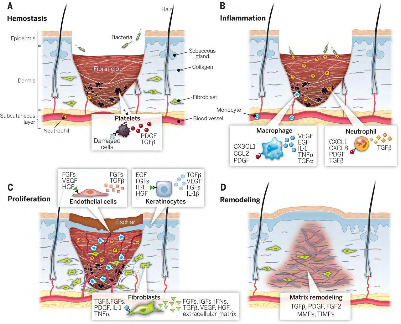
Figure 1: Stages of wound healing. It is classically divided into four stages: (A) hemostasis, (B) inflammation, (C) proliferation, and (D) remodeling. Each stage is characterized by key molecular and cellular events and is coordinated by a host of secreted factors that are recognized and released by the cells as a wounding response. Representative subsets of major factors are depicted. CCL2, chemokine (C-C motif) ligand 2; CX3CL1, chemokine (C-X3-C motif) ligand 1; CXCL1, chemokine (C-X-C motif) ligand 1; CXCL8, C-X-C Motif Chemokine Ligand 8; EGF, Epidermal growth factor; FGF2, basic fibroblast growth factor; FGFs, fibroblast growth factors; HGF, hepatocyte growth factor; IFNs, interferons; IGFs, insulin-like growth factors; IL-1, interleukin-1; IL-1β, interleukin-beta; MMPs, matrix metalloproteinases; PDGF, platelet-derived growth factor; PDGI, platelet- derived growth inhibitor; PGF2, Prostaglandin F2; TGFα, transforming growth factor-alpha; TGFβ, transforming growth factor- beta; TIMPs, tissue inhibitor of metalloproteinases; TNF-α, tumor necrosis factor alpha; VEGF, vascular endothelial growth factor (28).
The aim of wound treatment is to promote tissue repair and regeneration in a shorter time with adequate tissue perfusion and oxygenation, proper nutrition, and minimum undesired consequences (29).
Ethno-Medicinal Plants Used in Wound Healing
Ethno-medicinal plants are presumed to play a central role in remedies of wound healing since ancient times. A huge number of plants/plant extracts/decoctions or pastes are equally used by tribal and folklore traditions in Nepal, India, Bangladesh for the treatment of wounds and burns. It is important to note that about 80% of the Asian and African population still relies on the use of traditional medicine as primary health care [30]. However, the innovation of allopathic medicine retarded the utilization of ethno medicine. Due to various side effects of allopathic drugs, the uses of complementary and alternative medicines are gaining popularity for the treatment of wounds throughout the world [31]. These therapies have been proved cost-effective and efficacious for the treatment of diverse and difficult-healing wounds such as ulcers, burns, and infected wounds by stimulating the healing process and improving the strength of the new skin [32]. In this review, we elucidated the healing properties of various ethnomedicinal plants used in studies and outlined the information that might be useful to design the novel experiment for their scientific validation. Table 1 provides a general idea of the most important curative plants and their properties, with recognized effects on wound healing.
| Species/ plant’s name | Scheme | The extract used and formulation | Clinical properties | Wound model | Results | Refer ence |
|---|---|---|---|---|---|---|
| Achyranthe saspera | An ointment containing 5% methanol extract | Wound healing activity and antioxidant potential | Burn wound in a rat | It exhibited wound healing not only via wound contraction, fibroblast proliferation, epidermis formation, and collagen deposition, but also through the elevation of Na+, catalase, vitamin-C, and hydroxyl proline and the up-regulation of the expressions of matrix metalloproteinases (MMP-2 and MMP-9). | [33] | |
| Aloe vera | Blended fibrin/Aloe gel film | Wound healing | Streptozotocin- induced diabetic rats | The application enhanced wound healing by enhancing hydroxyproline, fibroblast, and collagen. | [34] | |
| Becium grandiflorum | 5% and 10% (w/w) ethanol leaf extract | Antibacterial, anti- inflammatory, and wound healing activity | Excision and incision wound models in mice | Ointments showed an increase in wound contraction, shorter epithelization time, and higher skin tensile strength along with considerable deposition of collagen, fibroblast proliferation, and vascularization. The plant extract also inhibited the inflammation. | [35] | |
| Buteamon osperma (Palas) | The alcoholic bark extract of Buteamono sperma | Cutaneous wound healing and antioxidant properties | Excision wound model of rat | The extract showed the increase in DNA, total protein, and collagen content of granulation tissues with more epithelialization and contraction. | [36] | |
| Caesalpinia mimosoides | Aqueous and ethanolic extract ointment 5% (w/w) of shoots and leaves | Antimicrobial, wound healing, and antioxidant activities | Circular excision and linear incision wound models in adult Wistar albino rats | Complete wound healing was noted with ethanol and aqueous extract on day 15. | [5] | |
| Calendula officinalis | n-hexanic and the ethanolic extracts from Calendula flowers | Wound healing | In vitro studies in human immortalized keratinocytes | Extracts influenced the inflammatory phase by activating the transcription factor NF-κB and by increasing chemokine IL-8, both at transcriptional and protein level. | [37] | |
| Calotropis procera | Topical application of 20 ml of 1.0% sterile solution of the latex of C. procera twice daily | Wound healing | Full-thickness excisional wounds of 8.0 mm diameter were inflicted in a guinea pig. | It resulted in wound area reduction by increasing collagen, DNA, protein synthesis, and epithelization. | [38] | |
| Carica papaya | Ethanol extract of seeds (50 mg/kg/ day) | Wound-healing activity, antimicrobial activity | Excision wound model in Sprague-Dawley rats | The papaya seed extract (1:1 ratio) showed 86% wound contraction on day 13. It also exhibited antimicrobial activity against Salmonella choleraesuis and staphylococcus aureus. | [39] | |
| Centella asiatica | Asiaticoside isolated from Centellaasiatica gum. Topical applications of 0.2% solution of Asiaticoside. | Healing activity | In-vivo study in guinea pig in punch wound model and in-vitro study in chick chorioallantoic membrane model | Asiaticoside produced a 56% increase in hydroxyproline, 57% increase in tensile strength, increased collagen content, and better epithelization. | [40] | |
| Daucu scarota | Paraffin-based cream containing 1%, 2%, and 4% w/w of ethanolic extract of root topically. | Wound healing activity | Excision wound model and incision wound model | It increased the hydroxyproline, 57% increase in tensile strength, increased collagen content,,t and better epithelization. | [12] | |
| Deloni xelata | Ethanolic extract (DSE) | Wound healing activity | Excision, incision, and dead space wound models | The complete wound contraction was found at 16 days. An increased expression of Col 1α (I) was observed in the wound tissue treated with DSE. | [10] | |
| Entadap haseoloides (L.) Merr. | Total Entadaphaseoloides (L.) Merr. tannins (TEPT) are used topically. | Wound healing activity | Excision wound model in rats | It promoted the wound shrinkage, rate and augmented the healing of infected wounds in rats with antimicrobial activity too. | [41] | |
| Globularia alypum | Methanolic extract | Burn wound healing process, inflammation, antibacterial and antioxidant activities | Burn wound of 177 mm2 area | Wound area contraction on day 12. | [42] | |
| Heliotropium indicum | Ethanolic extracts 10% w/v | Wound healing activity | Excision and incision wound models in rats | Complete healing of wounds was noted at 14 days. | [43] | |
| Hypericum patulum | 5% and 10% w/w ointment of methanol extract of leaves | Wound healing | Excision and an incision wound models in rats | Both concentrations of ointment enhanced wound healing. | [44] | |
| Jatrophaneo pauciflorapax | Two groups of treatment: Oral administration of latex (250, 500, and 750 mg/ kg) and topical administration of latex. | Antimicrobial, anti- inflammatory activities, and wound-healing | Anti- inflammatory activities in Wistar rats | It showed higher antioxidant activity, anti-inflammatory potential in oral administration of latex, and antimicrobial properties to promote wound healing. | [45] | |
| Lavandula aspic l. | Lavender oil | Antioxidant and wound healing activity | Excision wound model in rats | The biopsied skin showed fibrous connective tissue regeneration on day 14. | [46] | |
| Morinda citrifolia l. | Ethanolic extract of the leaf at a rate of 150 mg /kg/day orally | Wound healing activity | Excision and dead space wound models in rats | It enhanced wound contraction, decreased epithelialization time, and increased hydroxyproline content and histological characteristics. | [11] | |
| Musa sapientumvar. paradisiaca | Aqueous and methanolic extract of Musa sapientumb (100 mg/kg) orally. | Wound healing activity | Excision, Incision and dead space wound models in rats | Both extracts increased the levels of hydroxyproline, hexuronic acid, hexosamine, superoxide dismutase, reduced glutathione in granulation tissue, and decreased the wound area, scar area, and lipid peroxidation. | [47] | |
| Ocimum sanctum Linn | Alcoholic and aqueous extract 0f 400 mg/kg and 800 mg/kg body weight of each. | Antioxidant and wound healing effects | Incision, excision, and dead space wounds in Wistar albino rats | Both alcoholic and aqueous extract increased wound breaking strength, hydroxyproline, hexuronic acid, hexosamines, superoxide dismutase, catalase, reduced glutathione, and significantly decreased wound contraction and lipid peroxidation. | [48] | |
| Pereskia aculeate | 5 % of each methanol extract (me) and hexane fraction (hf) of leaves | Wound healing and anti- inflammatory potential | Excision wound | Hexane fraction followed by methanol extract markedly accelerated the wound closure. | [49] | |
| Plumbago zeylanicum | Ethanolic extracts 10% w/v | Wound healing activity | Excision and incision wound models in rats | Wound completely healed on day 18.4 | [43] | |
| Plumeria rubralinn | 0.5 % w/w Plumeriarubralinn (protease from the latex) ointment in the hydrophilic base was applied topically once a day | Inflammatory activities and wound-healing | Excision wound model in rats | It reduced the carrageenan-induced edema and resulted in 81% wound contraction at 16 days. | [50] | |
| Pongamia pinnata | Methanolic extracts of leaf | Wound healing, antimicrobial and antioxidant potential | Excision and an incision wound models in rats | Itdecreased the wound size (30 mm2) on day 16. | [51] | |
| Rhuschinensis | Hydroalcoholic 5% and 10% w/w leaf extract | Wound healing potential is also used to treat hemoptysis, inflammations, laryngitis, snakebite, stomachache, and fractures. | Incision wound, excision wound, and dead space wound | 10 % w/v extract had greater wound healing abilities than others but less than povidone- iodine. | [52] | |
| Sambucu sebulus | Alcohol preparation of powder of leaves and aerial parts of plants | Wound healing | Full-thickness excision wound model in rat | S. ebulus (2%) and its 2% combination enhanced wound healing. | [53] | |
| Stevia rebaudiana | Aqueous crude extract of leaves | Wound healing potential | Excision and incision wound model in mice | Stevia treated mice showed a significant reduction in the wound area, the faster rate of epithelialization with moderately higher hydroxyproline. | [54] | |
| Thevetiaper uviana | Leaves hexane extraction (lh) and fruit water extraction (fw) | Wound healing with antimicrobial, antioxidant, and anti- inflammatory potentials | Incision, excision, and dead space models in rats | Complete wound contraction was noted at 14 days. | [55] | |
| Wedelia chinensis | Ethanolic leaf extract | Wound healing activity | Excision, incision and dead space wound models in rats | Ethanolic extract retarded the period of epithelialization, increased the wound contraction, skin breaking strength, granulation tissue dry weight, and breaking strength. | [56] |
Table 1: shows the medicinal plants that are used for different models of the wound healing.
Areas of Interest for Researchers
In this world, nature gifted us a huge number of plants that are being used traditionally for medicinal purposes by various ethnic groups. Since ancient times, the plants, their extracts or paste have been consistently used to treat various types of wounds and tissue-related diseases. Further validation regarding the therapeutic uses of medicinal plants for healing wounds through scientific investigation seems imperative. The study on the underlying wound healing mechanism behind the use of medicinal plants could be a novel research area. Still, the several challenges such as identification, isolation, targeting, and mechanism of action of bioactive components of the medicinal plants are underexplored for developing the suitable herbal medicine for wound healing formulations. The combination of plant extracts with allopathic medicines may have synergistic potentials to promote wound healing capability and could be of recent research interest.
Conclusion
Wound healing is a complex process of the body self- defense mechanism. The topical or oral uses of herbal medicine result in augmenting the wound contraction, epithelization rate, granulation tissue dry weight, and its breaking strength, preventing infection with proper activities of different growth factors, and collagen, etc. Although traditional and complementary medicines were used as therapeutics in the ancient period, still some ethnic groups are using them regularly. The various clinical trials in animal models, as well as in-vitro studies, proved its potentiality for wound healing purposes. The ethnomedicinal plants also possess superior wound healing abilities even in diabetic wounds. Further molecular investigations on medicinal plants are imperative for the invention of novel drugs for wound healing with minimal side effects.
Conflict of Interest
The authors declare that there is no conflict of interests.
Acknowledgment
The authors are thankful to undergraduate students who helped in the search of the relevant research articles.
References
-
Pereira RF, Bártolo PJ (2016) Traditional Therapies for Skin Wound Healing. Adv Wound Care (New Rochelle) 5(5): 208-229.
-
Wild T, Rahbarnia A, Kellner M, Sobotka L, Eberlein T (2010) Basics in nutrition and wound healing. Nutrition 26(9): 862-866.
-
Mori HM, Kawanami H, Kawahata H, Aoki M (2016) Wound healing potential of lavender oil by acceleration of granulation and wound contraction through induction of TGF-β in a rat model. BMC Complement Altern Med 16: 144.
-
Akkol EK, Koca U, Peşin I, Yilmazer D, Toker G, et al. (2009) Exploring the wound healing activity of Arnebia densiflora (Nordm.) Ledeb by in vivo models. J Ethnopharmacol 124 (1): 137-141.
-
Bhat PB, Hegde S, Upadhya V, Hegde GR, Habbu PV, Mulgund GS (2016) Evaluation of wound healing property of Caesalpinia mimosoides Lam. J Ethnopharmacol 193: 712-724.
-
Deshmukh PT, Fernandes J, Atul A, Toppo E (2009) Wound healing activity of Calotropis gigantea root bark in rats. J Ethnopharmacol 125(1): 178-181.
-
Edwin S, Jarald EE, Deb L, Jain A, Kinger H, et al. (2008) Wound healing and antioxidant activity of Achyranthes aspera. Pharmaceutical Biology 46(12): 824-828.
-
El-Ferjani RM, Ahmad M, Dhiyaaldeen SM, Harun FW, Ibrahim MY, et al. (2016) In vivo Assessment of Antioxidant and Wound Healing Improvement of a New Schiff Base Derived Co (II) Complex in Rats. Scientific Reports 6(1): 1-12.
-
Kandhare AD, Alam J, Patil MVK, Sinha A, Bodhankar SL (2016) Wound healing potential of naringin ointment formulation via regulating the expression of inflammatory, apoptotic and growth mediators in experimental rats. Pharm Biol 54(3): 419-432.
-
Krishnappa P, Venkatarangaiah K, Venkatesh, ShimogaRajanna SK, KayattukandyBalan R (2016) Wound healing activity of Delonix elata stem bark extract and its isolated constituent quercetin-3- rhamnopyranosyl-(1-6) glucopyranoside in rats. J Pharm Anal 6(6): 389-395.
-
Nayak BS, Sandiford S, Maxwell A (2009) Evaluation of the wound-healing activity of ethanolic extract of Morinda citrifolia L. leaf. Evidence-based Complement. Altern Med 6(3): 351-356.
-
Patil MVK, Kandhare AD, Bhise SD (2012) Pharmacological evaluation of ethanolic extract of Daucus carota Linn root formulated cream on wound healing using excision and incision wound model. Asian Pac J Trop Biomed 2(2): S646-S655.
-
Saratha V, Subramanian S, Sivakumar S (2010) Evaluation of wound healing potential of calotropis igantea latex studied on excision wounds in experimental rats. Med Che Res 19: 936-947.
-
Wang L, Qin W, Zhou Y, Chen B, Zhao X, et al. 2017) Transforming growth factor β plays an important role in enhancing wound healing by topical application of Povidone-iodine. Sci Rep 7(1): 991.
-
Subramanian S, Kumar DS, Arulselvan P (2006) Wound Healing Potential of Aloe vera Leaf Gel Studied in Experimental Rabbits. Asian Journal of Biochemistry 1(2): 178-185.
-
Gurtner GC, Werner S, Barrandon Y, Longaker MT (2008) Wound repair and regeneration. Nature 453: 314-321.
-
Schreml S, Szeimies RM, Prantl L, Landthaler M, Babilas P (2010) Wound healing in the 21st century. J Am Acad Dermatol 63(5): 866-881.
-
Barrientos S, Stojadinovic O, Golinko MS, Brem H, Tomic Canic M (2008) Growth factors and cytokines in wound healing. Wound Repair Regen 16(5): 585-601.
-
Kolaczkowska E, Kubes P (2013) Neutrophil recruitment and function in health and inflammation. Nat Rev Immunol 13 (3): 159-175.
-
Ridiandries A, Tan JTM, Bursill CA (2018) The Role of Chemokines in Wound Healing. Int J Mol Sci 19 (10): 3217.
-
Behm B, Babilas P, Landthaler M, Schreml S (2012) Cytokines, chemokines and growth factors in wound healing. J Eur Acad Dermatology Venereol 26(7): 812- 820.
-
Werner S, Grose R (2003) Regulation of wound healing by growth factors and cytokines. Physiol Rev 83(3): 835- 870.
-
Clark RAF, Nielsen LD, Welch MP, McPherson JM (1995) Collagen matrices attenuate the collagen-synthetic response of cultured fibroblasts to TGF-β. J Cell Sci 108 (3): 1251-1261.
-
Desmouliere A, Geinoz A, Gabbiani F, Gabbiani G (1993) Transforming growth factor-β1 induces α-smooth muscle actin expression in granulation tissue myofibroblasts and in quiescent and growing cultured fibroblasts. J Cell Biol 122 (1): 103-111.
-
Singer AJ, Clark RA (1999) Cutaneous wound healing. N Engl J Med 341(10): 738-46.
-
Lever E, Sheer D (2010) The role of nuclear organization in cancer. J Pathol 220 (2): 114-125.
-
Shih B, Garside E, McGrouther DA, Bayat A (2010) Molecular dissection of abnormal wound healing processes resulting in keloid disease. Wound Repair Regen 18: 139-153.
-
Sun BK, Siprashvili Z, Khavari PA (2014) Advances in skin grafting and treatment of cutaneous wounds. Science 346(6212): 941-945.
-
Pierce GF, Mustoe T A (1995) Pharmacologic enhancement of wound healing. Annu Rev Med 46: 467– 81.
-
Oyebode O, Kandala NB, Chilton PJ, Lilford RJ (2016) Use of traditional medicine in middle-income countries: a WHO-SAGE study. Health Policy Plan 31(8): 984-991.
-
Sabale P, Bhimani B, Prajapati C,Sabalea V (2012) An overview of medicinal plants as wound healers. J Appl Pharm Sci 2(11): 143-150.
-
Maver T, Maver U, Kleinschek SK, Smrke DM, Kreft S (2015) A review of herbal medicines in wound healing. Int J Dermatol 54 (7): 740-751.
-
Barua CC, Talukdar A, Begum SA, Pathak DC, Sarma DK, et al. (2012) In vivo wound-healing efficacy and antioxidant activity of Achyranthes aspera in experimental burns. Pharm Biol 50 (7): 892-899.
-
Inpanya P, Faikrua A, Ounaroon A, Sittichokechaiwut A, Viyoch J (2012) Effects of the blended fibroin/aloe gel film on wound healing in streptozotocin-induced diabetic rats. Biomed Mater 7 (3): 035008.
-
Beshir K (2017) Evaluation of Wound Healing Activity of 70% Ethanol Leaf Extract of Becium grandiflorum Lam. (Lamiaceae) in Mice. Ethiopian Pharmaceutical Journal 32(2): 117-130.
-
Sumitra M, Manikandan P, Suguna L (2005) Efficacy of Butea monosperma on dermal wound healing in rats. Int. J Biochem Cell Biol 37(3): 566-573.
-
Nicolaus C, Junghanns S, Hartmann A, Murillo R Ganzera M, et al. (2017) In vitro studies to evaluate the wound healing properties of Calendula officinalis extracts. J Ethnopharmacol 196: 94-103.
-
Rasik AM, Raghubir R, Gupta A, Shukla A, Dubey MP, Srivastava S, Jain HK, Kulshrestha DK (1999) Healing potential of Calotropis procera on dermal wounds in Guinea pigs. J Ethnopharmacol 68 (1-3): 261-266.
-
Nayak BS, Ramdeen R, Adogwa A, Ramsubhag A, Marshall JR (2012) Wound-healing potential of an ethanol extract of Carica papaya (Caricaceae) seeds. Int Wound J 9 (6): 650-655.
-
Shukla A, Rasik AM, Jain GK, Shankar R, Kulshrestha, DK, et al. (1999) In vitro and in vivo wound healing activity of asiaticoside isolated from Centella asiatica. J Ethnopharmacol 65(1): 1-11.
-
Su X, Liu X, Wang S, Li B, Pan T, et al. (2017) Wound- healing promoting effect of total tannins from Entada phaseoloides (L.) Merr in rats. Burns 43(4): 830-838.
-
Ghlissi Z, Kallel R, Sila A, Harrabi B, Atheymen R, et al. (2016) Globularia alypum methanolic extract improves burn wound healing process and inflammation in rats and possesses antibacterial and antioxidant activities. Biomed Pharmacother 84: 1488-1495.
-
Reddy JS, Rao PR, Reddy MS (2002) Wound healing effects of Heliotropium indicum, Plumbago zeylanicum and Acalypha indica in rats. J Ethnopharmacol 79 (2): 249-251.
-
Mukherjee PK, Verpoorte R, Suresh B (2000) Evaluation of in-vivo wound healing activity of Hypericum patulum (Family: Hypericaceae) leaf extract on different wound models in rats. J Ethnopharmacol 70(3): 315-321.
-
Hernandez-Hernandez AB, Alarcon-Aguilar FJ, Almanza- Perez JC, Nieto-Yañez O, Olivares-Sanchez JM, et al. (2017) Antimicrobial and anti-inflammatory activities, wound- healing effectiveness and chemical characterization of the latex of Jatropha neopauciflora Pax. J Ethnopharmacol 204: 1-7.
-
Ben Djemaa FG, Bellassoued K, Zouari S, El Feki A, Ammar E (2016) Antioxidant and wound healing activity of Lavandula aspic L. ointment. J Tissue Viability 25 (4): 193-200.
-
Agarwal PK, Singh A, Gaurav K, Goel S, Khanna HD, et al. (2009) Evaluation of wound healing activity of extracts of plantain banana (Musa sapientum var. paradisiaca) in rats. Indian J Exp Biol 47 (1): 32–40.
-
Shetty S, Udupa S, Udupa L (2008) Evaluation of antioxidant and wound healing effects of alcoholic and aqueous extract of Ocimum sanctum Linn in rats. Evidence-based Complement Altern Med 5(1): 95-101.
-
Pinto NC, Cassini-Vieira P, Souza-Fagundes EM, Barcelos LS, Castañon MC, et al. (2016) Pereskia aculeata Miller leaves accelerate excisional wound healing in mice. J Ethnopharmacol 194: 131-136.
-
Chanda I, Sarma U, Basu SK, Lahkar M, Dutta SK (2011) A Protease Isolated from the Latex of Plumeria rubra Linn ( Apocynaceae ) 2 : Anti-inflammatory and Wound- Healing Activities. Tropical Journal of Pharmaceutical Research 10(6): 755-760.
-
Dwivedi D, Dwivedi M, Malviya S, Singh V (2017) Evaluation of wound healing, anti-microbial and antioxidant potential of Pongamia pinnata in wistar rats. Journal of Traditional and Complementary Medicine 7(1): 79-85.
-
Haloi P, Sedhain A, Roy K (2016) Wound healing potential of the hydroalcoholic leaf extract of Rhuschinensis mill. The Pharm student 27: 10-21.
-
Babaei E, Asghari MH, Mehdikhani F, Moloudizargari M, Ghobadi E, Pouya SRH (2017) The healing effects of herbal preparations from Sambucusebulus and Urticadioica in full-thickness wound models. Asian Pac J Trop Biomed 7(5): 421-427.
-
Das K (2013) Wound healing potential of aqueous crude extract of Stevia rebaudiana in mice. Brazilian J Pharmacogn 23(2): 351-357.
-
Rahman N, Rahman H, Haris M, Mahmood R (2017) Wound healing potentials of Thevetia peruviana: Antioxidants and inflammatory markers criteria. J Tradit Complement Med 7(4): 519-525.
-
Verma N, Khosa RL, Garg VK (2008) Wound healing activity of Wedelia chinesis leaves. Pharmacologyonline 2: 139-145.
- Mitochondrial Bio-Logistics: Steering Co-Enzyme Q10 and Lycopene Synergies within the Science 4.0 Bio-OS Framework
- Hymenoptera Specimens from the Caño Negro Wetland, of the National Museum Collection, Costa Rica
- Science 4.0: Comprehensive Architecture of the Biological Operating System (Bio-OS) A Framework for Systemic Resilience and Industrialized Bio-Governance
- Rabbit on, or Hare Back? Understanding Climate Change
- Clinical Validation of Science 4.0: Flow Steering and Epigenetic Drift Inversion on a 76-Year-Old Hybrid System
- Seeds Planted by another Mind