Prevalence of Gastrointestinal Endoparasites in Captive Wild Animals at Aurangabad Municipal Corporation Zoo, Maharashtra, India
The present work was conducted on captive wild animals to study the prevalence of gastrointestinal endoparasites during three seasons (winter, summer and monsoon). In total 126 (45 Carnivores, 24 Herbivores, 1 Omnivore, 21 Birds, and 33 Reptiles) feacal samples were examined out of which 54 (24 Carnivores, 18 Herbivores, 2 Omnivore, 10 Birds, and 7 Reptiles) were positive for one or more parasites with prevalence of 48.41%. The study revealed statistically significant difference in the prevalence of endoparasites in winter (61.90%) as compared to summer (38.09%) and monsoon (45.23%). The prevalence of GI endoparasites was 53.33 % in carnivores (winter 66.66%, summer 46.66%, monsoon 46.66%), 75% in herbivores (winter 87.5%, summer 62.5%, monsoon 75%), 66.66% in omnivores (winter 100%, summer 100%, monsoon 0%), 47.61% in birds (winter 71.42%, summer 28.57%, monsoon 42.85%) and 21.21% in reptiles (winter 27.27%, summer 9.09%, monsoon 27.27%). Common GI endoparasites in carnivores were Toxocara spp., Strongyle spp., Trichuris spp., Ascaris spp. and Eimeria spp., in herbivores Strongyle spp., Schistosoma spp. and Eimeria spp., and in birds Ascaris spp. and Eimeria spp. The data obtained in this work could be used for implementation of effective management strategies against gastrointestinal endoparasites of various captive wild animal species.
Introduction
Wild animals are displayed in zoological gardens for aesthetic, recreational, educational, and conservation reasons. Parks and zoological gardens play a significant role in species conservation in many regions of the world. Parasitic infections in captive wild animals is a big issue leading to health implications and sometimes causing even death [1].
Wild animals in nature have broad habitats travelling over vast areas and, as a result due to minimal exposure have low genetic resistance to parasites. Animals at zoological parks are constantly stressed as a result of captivity, despite care and supervision, and are susceptible to parasite illnesses when they get exposed to parasites leading to diseases that can pose a serious threat to endangered species and inflicting sudden and unexpected local population decreases [2]. Endoparasite development in zoo animals is influenced by factors such as crowding, hygiene, and diet [3]. Therefore, identification of the parasites is essential for successful parasitic infection treatment and management in captive animals. Gastrointestinal parasite surveillance and control measures based on accurate diagnosis, successful treatment, and proper prophylaxis will undoubtedly aid in correcting the health status of captive wild animals. Due to changes in the host’s physiology and basal metabolic rate as a result of changing seasonal variables like temperature and photoperiods parasites show seasonal variation in infection rates [4], therefore, warranting a thorough investigation of diverse species of zoo-housed animals to determine the true prevalence for implementing better control and management techniques [5]. For effective management of captive wild animal health and public health issues, this study was aimed to analyse and describe the gastrointestinal parasites profile among animals kept in the Aurangabad Municipal Corporation Zoo, Maharashtra, India for implementation of effective management strategies against these parasites.
Materials and Methods
Sampling Strategy and Captive Wild Animals Involved in the Study
The present study was undertaken on captive wild animals of Siddharth Garden Aurangabad Municipal Corporation Zoo, Maharashtra to study the epidemiology of endoparasites affecting zoo animals and birds. To evaluate the prevalence of gastrointestinal parasites individual/ pooled faecal sample were collected from different species of captive wild animals (n=42) during winter, summer and monsoon seasons for a period of ten months from January to October, 2021.
Collection of Feacal Samples
Fresh faeces (0-12h) based on colour and moisture content of the faeces will be collected. The surface portion of the faeces were removed and the interior portion were collected in sterile whirl pack polythene bags using sterile spatulas for each sample. The samples were stored at 4°C for 48-72 hours before being transported to the laboratory for further processing.
Processing of Faecal Samples
The faecal samples were subjected to detailed conventional parasitological analysis for presence of parasitic eggs/ oocysts by direct smear examination, standard floatation and sedimentation techniques as given by Soulsby, et al. [6]. Also faecal samples were processed for identification and antimicrobial sensitivity testing of prevalent bacterial/ enteric pathogens including E. coli, Salmonella, Klebsiella etc using standard microbial procedures.
Direct Smear Examination: A pinch of faecal sample were placed on one end of a slide and after mixing with a drop of water, were spread on slide, covered with cover slip, and examined directly at low power (10X) followed by high power (40X) objectives of the microscope. At least three slides from different parts of the faecal samples were examined.
Floatation Concentration Technique: About 2g faecal sample were taken into a pestle and mortar and a suspension of the sample strainable with a sieve were made by adding water, followed by complete mixing. The strained material was taken into a beaker and well mixed with Sheather’s sugar solution avoiding air bubble formation. The suspension was then transferred into a centrifuge tube and filled up to the brim until a convex meniscus formed. A cover glass was placed on it avoiding air bubble formation. After about 30 minutes, the cover glass was gently lifted in a horizontal position and mounted on a glass slide and focused under microscope, initially under low power (10X), followed by high power (40X) for detailed study.
Sedimentation Technique: About 2 g of faecal sample was triturated with water in a pestle and mortar and then filtered through a sieve into a beaker. Contents was allowed to settle for 20-30 minutes after which the supernatant was discarded and sediment was examined on a glass slide for the presence of parasitic eggs under the low power (10X) and high power (40X) of the microscope.
Sporulation of Oocysts: The faecal samples found positive for coccidian oocysts by direct and floatation concentration techniques was cultured for sporulation of oocysts. The positive faecal sample was triturated in Potassium dichromate solution (2.5%) in a pestle and mortar and then transferred to a petri dish. The petri dish was filled up to the mark covering the faecal material and was left as such at room temperature or kept in incubator (preferably at 27°C) with periodic aeration and addition of Potassium dichromate solution (2.5%). The material was regularly checked for sporulation of the oocysts by taking a drop of the suspension on to a glass slide (or by floatation concentration method) and examined it under the low power (10X) and later high power (40X) of the microscope to observe the detailed morphology.
Statistical Analysis
The data was analysed using Statistical software program (SPSS for Windows, Version 19.0, USA) and to know the effect of season on the prevalence of GI endoparasites in the captive wild animals the Chi-square test used.
Results
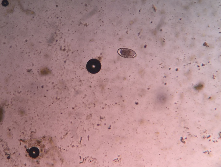
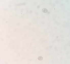
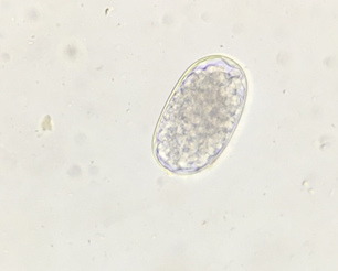
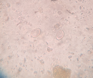
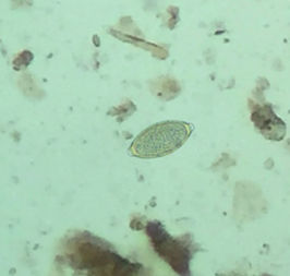
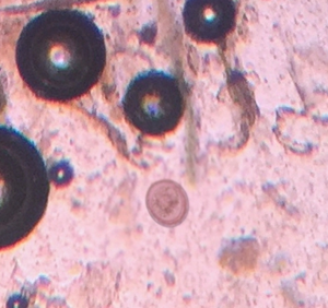
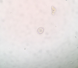
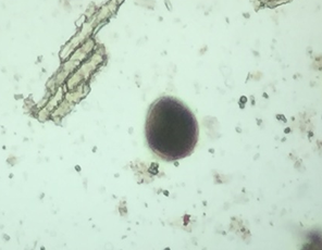
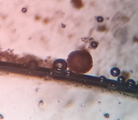
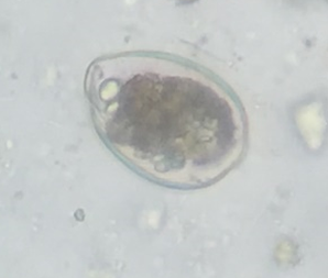
In this study the prevalence of GI endoparasites was observed for ten months during three seasons (winter, summer and monsoon). During this period 126 (45 Carnivores, 24 Herbivores, 1 Omnivore, 21 Birds and 33 Reptiles) faecal samples were examined out of which 54 (24 Carnivores, 18 Herbivores, 2 Omnivore, 10 Birds, and 7 Reptiles) were found positive for one or more parasitic infection with an overall prevalence of 48.41% (winter 60.90 %, summer 38.09%, monsoon 45.23%) (Table 1). Out of 24 herbivores 18 were positive for one or more parasitic infection revealing the prevalence of 75% (Table 1). Herbivores were found infected with Strongyle spp., Schistosoma spp. and Eimeria spp. (Table 3 & Figures 1,3,6). The prevalence of GI endoparasites in carnivores (53.33%) including Tiger, Leopard, Jackal and Indian Civet cat was lower as compared to herbivores (75%) and omnivores (66.66%). The most common endoparasites found in carnivores were Toxocara spp., Strongyle spp., Trichuris spp., Ascaris spp. and Eimeria spp. (Table 2 & Figures 2,5,7,10-12). Pooled faecal sample of monkeys examined during winter and summer revealed Eimeria spp. oocysts (Figure 8) while as did not reveal any oocyst during monsoon season. The study revealed 47.61% prevalence of helminthic parasites in birds (Table 1). The study revealed Ascaridia spp. and Eimeria spp. (Table 4 & Figure 9) as most common helminthes observed in birds. The study revealed 21.21% prevalence of GI parasites in captive reptiles (Table 1). Most common GI parasites observed were Strongyle spp. and Eimeria spp. in snakes (Table 5).
| Animals | Winter | Summer | Monsoon | Total |
|---|---|---|---|---|
| Total Carnivores | 15 | 15 | 15 | 45 |
| Positive | 10 | 7 | 7 | 24 |
| Prevalence (%) | 66.66 | 46.66 | 46.66 | 53.3 |
| Total Herbivores | 8 | 8 | 8 | 24 |
| Positive | 7 | 5 | 6 | 18 |
| Prevalence (%) | 87.5 | 62.5 | 75 | 75 |
| Total Omnivores | 1 | 1 | 1 | 3 |
| Positive | 1 | 1 | 0 | 2 |
| Prevalence (%) | 100 | 100 | 0 | 66.7 |
| Total Birds | 7 | 7 | 7 | 21 |
| Positive | 5 | 2 | 3 | 10 |
| Prevalence (%) | 71.42 | 28.57 | 42.85 | 47.6 |
| Total Reptiles | 11 | 11 | 11 | 33 |
| Positive | 3 | 1 | 3 | 7 |
| Prevalance (%) | 27.27 | 9.09 | 27.27 | 21.2 |
| Overall Prevalence (%) | ##### | 38.09% | 45.23% | ### |
| (26/42) | (16/42) | (19/42) | (61/126) | |
| χ2 | 2.977* | |||
| Sr No. | Common name | Winter | Summer | Monsoon |
| A | Tiger | |||
| 1 | Bengal Tiger (Sidhharth) | Toxocaraspp. | ND | Strongyle spp. |
| 2 | Bengal Tiger (Arjun) | Strongyle spp. | ND | Strongyle spp. |
| 3 | Bengal Tiger (Kush) | ND | Strongyle spp. | ND |
| 4 | White Bengal Tiger (Veer) | ND | ND | Trichurisspp. |
| 5 | Bengal Tiger (Samrudhi) | Toxocaraspp., Strongyle spp. | Trichurisspp., Strongyle spp. | Toxocaraspp., Strongyle spp. |
| 6 | Bengal Tiger (Bhakti) | ND | ND | ND |
| 7 | White Bengal Tiger (Pragati) | Trichurisspp., Toxocaraspp. | Strongylespp., Toxocaraspp. | Strongylespp., Toxocara spp. |
| 8 | White Bengal Tiger (Arpita) | Trichuris spp. | ND | ND |
| 9 | Bengal Tiger (Devika) | Toxocaraspp. | Strongyle spp. | ND |
| 10 | Bengal Tiger (Cub’s) | ND | ND | Toxocaraspp. |
| B. | Leopard | |||
| 1 | Leopard 1 | Ascaris spp. | Toxocaraspp. | Strongyle spp. |
| 2 | Leopard 2 | Toxocaraspp. | ND | ND |
| C. | Jackal | |||
| 1 | Jackal (mass) | Eimeria spp. | Trichuris spp. | ND |
| 2 | Jackal | ND | ND | ND |
| D. | Indian Civetcat | |||
| 1 | Indian Civetcat | Eimeria spp. | Toxocaraspp. | ND |
Table 1: Prevalence of GI parasites in various captive animals and birds.
*(χ2 = 2.977; 02df; P<0.05). Table 1: Prevalence of GI parasites in various captive animals and birds.
*ND-Sample processed and nothing detected. Table 2: Break down of various parasitic eggs/ oocysts recovered from different Carnivores.
| Sr No. | Common name | Winter | Summer | Monsoon |
|---|---|---|---|---|
| A | Sambar Deer | |||
| 1 | Sambar Deer (Pooled) | Strongyle spp. | Strongyle spp. | Strongyle spp. |
| 2 | Sambar Deer Male | ND | ND | ND |
| 3 | Sambar Deer Female | Strongyle spp. | ND | Strongyle spp. |
| B. | Blackbuck | |||
| 1 | Blackbuck | Strongyle spp. | Strongyle spp. | Strongyle spp. |
| C. | Spotted Deer | |||
| 1 | Spotted Deer | Strongyle spp. | Eimeriaspp. | Strongyle spp. |
| D. | Indian Porcupine | |||
| 1 | Male Porcupine | Schistosoma spp. | ND | ND |
| 2 | Female Porcupine | Strongyle spp. | Strongyle spp. | ND |
| E. | Nilgai | |||
| 1 | Nilgai | Strongyle spp. | Strongyle spp. | Strongyle spp. |
| S. No. | Common name | Winter | Summer | Monsoon |
| A | Birds | |||
| 1 | Emu | Ascaris spp. | Ascaris spp. | Ascaris spp. |
| 2 | Indian Peafowl | Eimeria spp. | ND | Eimeria spp. |
| 3 | Painted Storck | ND | Eimeria spp. | ND |
| 4 | Little Egret | Ascaris spp. | ND | ND |
| 5 | Spoon Bill | Eimeria spp. | ND | ND |
| 6 | White Necked Storck | ND | ND | Eimeria spp. |
| 7 | Grey Heron | Eimeria spp. | ND |
Table 2: Break down of parasitic eggs/ oocysts recovered from different herbivores.
*ND- Sample processed and nothing detected. Table 3: Break down of parasitic eggs/ oocysts recovered from different herbivores.
*ND- Sample processed and nothing detected. Table 4: Break down of parasitic eggs/ oocysts recovered from different captive birds.
| Sr No. | Common name | Seasons | ||
|---|---|---|---|---|
| A | Snakes | Winter | Summer | Monsoon |
| 1 | Indian Rock Python | ND | ND | Strongyle spp. |
| 2 | Common Trinket | ND | ND | ND |
| 3 | Banded Racer | Strongyle spp. | ND | Eimeria spp. |
| 4 | Grass Snake | ND | ND | ND |
| 5 | Earth Boa | ND | ND | ND |
| 6 | Indian Rat Snake | Strongyle spp. | Strongyle spp. | ND |
| 7 | Common krait | ND | ND | ND |
| 8 | Indian Cobra | ND | ND | ND |
| 9 | Russll’s Viper | Eimeria spp. | ND | Strongyle spp. |
| F. | Crocodile | ND | ND | ND |
| 1 | Male Crocodile | ND | ND | ND |
| 2 | Female Crocodile | ND | ND | ND |
Table 3: Break down of parasitic eggs/ oocysts recovered from different captive Reptiles.
*ND- Sample processed and nothing detected. Table 5: Break down of parasitic eggs/ oocysts recovered from different captive Reptiles.
The study revealed that overall prevalence of endoparasites was high in winter (61.90%) as compared to summer (38.09%) and monsoon (45.23%) (Table 1) and the variation in prevalence between different seasons was found statistically significant (χ2 = 2.977; 02df; P<0.05). The prevalence of GI endoparasites recorded in carnivores was 53.33 % (winter 66.66%, summer 46.66%, monsoon 46.66%), in herbivores 75 % (winter 87.5%, summer 62.5%, monsoon 75%) and in omnivores 66.66% (winter 100%, summer 100%, monsoon 0%), in birds 47.61% (winter 71.42%, summer 28.57%, monsoon 42.85%) and in reptiles 21.21% (winter 27.27%, summer 9.09%, monsoon 27.27%).
Discussion
Overall prevalence of GI endoparasites in this study was observed as 48.41% which corroborate with research findings of other researchers like Reddy, et al. [7]; Chakraborty, et al. [8]; Modi, et al. [9] and Thawait, et al. [10] who reported GI endoparasite prevalence of 42.4%, 40.4%, 48.1% and 46.2% respectively. The study on prevalence of GI parasites has been conducted in various zoos and national parks throughout the world by different researchers like Mir, et al. [11]; Maske, et al. [12]; Opara, et al. [13]; Parsani, et al. [14; Thawait, et al. [10] and Rahman, et al. [15]. The prevalence of GI endoparasites observed in our study was comparatively lower than the previous findings of some researchers like Cordon, et al. [16]; Thawait, et al. [10]; Opara, et al. [13]; Rahman, et al. [15] and Varadharajan, et al. [17] who reported GI endoparasite prevalence of 72.5, 68.05, 76.6%, 76.9% and 68.36 respectively.
In this study we observed that herbivores kept in herds including Sambar deer, Black buck, Spotted deer, Nilgai
showed higher prevalence in comparison to the omnivores and individually enclosed carnivores including Tiger, Leopard, Jackal, Indian Civetcat. Similar findings were also observed by some researchers like Vardharajan, et al. [17] who reported higher prevalence of helminthic infection in herbivores (71.62%) than the omnivores (65.9%) and Rehman, et al. [15] reported prevalence of 76.9% in herbivores. Usually overcrowding in herd animals, competition for feed and water results in stress and decreased immunity, leading to more vulnerability to parasitic infections [18, 19, 20]. Contrary to our findings lower prevalence in herbivores was observed by others like Thawait, et al. [10]; Varadharajan, et al. [21]; Singh, et al. [22]; Thawait, et al. [23]; Mudgil, et al. [18] and Mir, et al. [11] who reported prevalence of 45.6%, 67.47%, 25.71%, 45.68%, 23.30% and 68% respectively.
Lower prevalence in carnivores in comparison to herbivores and omnivores could be contributed to their individual confinement and good managemental practices. Singh, et al. [22] reported 58.68% prevalence in carnivores at Mahendra Choudhury Zoological Park, Chhatbir, Panjab and Mahali, et al. [24] reported 60.52% prevalence in carnivores of Nandankanan Zoo, Bhubaneswar, Odisha. Some researchers observed lower prevalence in carnivores than our study like Thawait, et al. [10]; Nasiri, et al. [25], Ramadevi, et al. [26] who reported prevalence as 37.24%, 13.88%, 23.59% respectively.
Relatively higher prevalence in omnivores could be contributed to small sample size in this study. Varadharajan, et al. [21] and Vardharajan, et al. [17] reported similar prevalence of 65.35% and 65.9% in omnivores respectively. Singh, et al. [20] and Mudgil, et al. [18] reported a lower GI parasitic prevalence of 29.02% and 6.85% respectively in omnivores while as Arunachalam, et al. [27] reported higher GI parasitic prevalence of 43% in Rhesus Macaque.
In contrast to our finding of 47.61% prevalence of helminthic parasites in birds Parasani, et al. [14]; Lim, et al. [28] and Fagiolini, et al. [29] reported higher prevalence of 60.7%, 56.3% and 61.5% respectively while as Hoque, et al. [30], Nasiri, et al. [31] and Ramadevi, et al. [26] reported lower prevalence of 20%, 4.81 and 9.09% respectively. Moudgil, et al. [18] also studied the prevalence of GIT parasitic infections in zoo-housed birds of various zoological/deer parks and an aviary of Punjab, India screening 1273 samples from the birds of the MC Zoological Park, BirMotibagh Deer Park Patiala, Patiala aviary, BirTalab Deer Park Bathinda and Tiger safari Ludhiana showing an overall GIT parasitic burden of 37.52 %, 25.54%, 37.50%, 45.39%, and 67.64% respectively. The finding of predominance of Ascaridia spp. and Eimeria spp. in birds could be attributed to direct life cycle of these parasites [14]. The protozoan infection mainly involved coccidian infection of Eimeria spp. a finding also reported by Morrondo, et al. [32] and Mudgil, et al. [18]. The study revealed lower prevalence (21.21%) of GI parasites in captive reptiles. Akhila, et al. [33] reported 71.4% prevalence of GI parasites in captive snakes of Kerala. Chaiyabutr and Chanhome, et al. [34] reported 75% prevalence of GI parasites in the snake farm of the Queen Saovabha Memorial Institute. Nasiri, et al. [25] reported prevalence of 47.12% in Iranian snakes.
The variation in prevalence between different seasons was found statistically significant (χ2 = 2.977; 02df; P<0.05). Some researchers also studied the seasonal prevalence in wild animals and reported different findings than our study like Mudgil, et al. [18] who reported monsoon season prevalence of 37.73% and 53.12% in animals and birds of MC Zoological Park, Chhatbir, Panjab and in the animals and birds of BirMotibagh Deer Park, Patiala respectively. Mahali, et al. [24] also studied the prevalence in the carnivores of Nandankanan Zoo during three seasons (Rainy, winter and summer) and reported higher incidence during rainy season (63.51%), as compared to summer (62.96%) and winter seasons (54.29%). High prevalence of GI endoparasites in winter observed in this study could be due to lack of deworming after monsoon season. Lower prevalence in summer as compared to winter season were also observed by other researchers like Modi, et al. [9] and Kumar, et al. [35, 36, 37, 38, 39, 40].
Conclusion
The study revealed an overall GI endoparasite prevalence of 48.41% and statistically significant variation in the prevalence between different seasons (winter 60.90 %, summer 38.09%, monsoon 45.23%). The prevalence of GI endoparasites observed in carnivores, herbivores, omnivores, birds and reptiles was 53.33 %, 75 %, 66.66%, 47.61% and 21.21% respectively. The data obtained in this work could be used for implementation of effective control and management strategies against gastrointestinal endoparasites of various captive wild animal species to minimize negative health impacts and consequences of parasitism in captive wild animals.
Acknowledgment
Authors are highly thankful to Chief Wildlife Warden, Maharashtra for giving the necessary permission and help to undertake this investigation.
• Declaration of interest statement The authors report no conflict of interest.
References
-
Rao AT, Acharjyo LN (1984) Diagnosis and classification of common diseases of captive animals at Nandankana Zoo in Orissa (India). Indian J Anim. H Dec 23(2): 147- 157.
-
Muoria PK, Muruthi P, Rubenstein D, Oguge NO, Munene E (2005) Cross-sectional survey of gastro-intestinal parasites of Grevy’s zebras in southern Samburu, Kenya. Afr J Ecol 43(3): 392-395.
-
Malan FS, Horak IG, Vos V, Van Wik JA (1997) Wildlife parasites: lessons for parasites control in livestock. Vet Parasitol 71(2-3): 137-153.
-
Patra A, Min SK, Seong MG (2020) Climate variability impacts on global extreme wave heights: seasonal assessment using satellite data and ERA5 reanalysis. Journal of Geophysical Research: Oceans 125(12): e2020JC016754.
-
Papini R, Girivetto, Marangi M, Mancianti F, Giangaspero A (2012) Endoparasite infections in pet and zoo birds in Italy. The scientific world journal 2012(1): 253127.
-
Soulsby JL (1968) Helminths, arthropods and protozoa of domesticated animals. In: 6th (Edn.), Helminths, arthropods and protozoa of domesticated animals, pp: 824
-
Reddy JNR, Jagannath MS, D’Souza PE, Abdul RS (1992) Prevalence of gastrointestinal parasites in wild mammals and captive birds at Bannerghata National Park, Bangalore, India. Indian Journal of Animal Sciences 62(1): 1046-1048.
-
Chakraborty A, Islam S (1996) A survey of gastrointestinal parasitic infections in some free-living herbivores in the Kaziranga National Park. Zoo’s Print Journal 16(1): 1-3.
-
Modi GS, Prasad BN, Sinha PK (1997) Effect of age on the prevalence of intestinal parasitism among zoo animals in Bihar. Indian Veterinary Journal 74(4): 351-353.
-
Thawait VK, Maiti SK, Dixit AA (2014) Prevalence of gastro-intestinal parasites in captive wild animals of Nandan Van Zoo, Raipur, Chhattisgarh. Veterinary World 7(7): 1-4.
-
Mir AQ, Dua K, Singla LD, Sharma S, Singh MP (2016) Prevalence of parasitic infection in captive wild animals in BirMotiBagh mini zoo (Deer Park), Patiala, Punjab. Veterinary World 9(6): 540-543.
-
Maske DK, Bhilegaonkar NG, Sardey MR (1990) Prevalence of parasitic infections in domestic animals at Nagpur (Maharashtra). Journal of Veterinary Parasitology 4(2): 23-25.
-
Opara M, Osuji C, Opara J (2010) Gastrointestinal parasitism in captive animals at the zoological garden, NekedeOwerri, Southeast Nigeria. Ostrich 2(5): 21-28.
-
Parsani HR, Momin RR, Maradia MG, Singh V (2001) A survey of gastrointestinal parasites of captive animals at Rajkot municipal corporation zoo, Rajkot, Gujarat. Zoo’s Print Journal 16(10): 604-606.
-
Rahman SM, Dey AR, Kundu UK, Begum N (2014) Investigation of gastrointestinal parasites of herbivores at Dhaka National Zoological Garden of Bangladesh. Journal of the Bangladesh Agricultural University 12(1): 79-85.
-
Cordon PG, HitosPrados A, Romero D, Moreno MS, Pontes A, et al. (2008) Intestinal parasitism in the animals of the zoological garden “Pe˜naEscrita” (Almunecar, Spain). Veterinary Parasitology 156(3-4): 302-309.
-
Varadharajan A, Subramanian H (2003) Influence of age on the prevalence of parasitic infections among wild mammals in Thrissur Zoo, Thrissur, Kerala. Zoos’ Print J 18(4): 1065-1066.
-
Moudgil AD, Singla LD, Singh MP (2020) Seasonal variation in gastrointestinal parasitism of zoo-housed birds of Punjab, India. Biological Rhythm Research 51(7): 1075-1086.
-
Dhoot VM, Upadhye SV, Kolte SW (2002) Prevalence of parasitism in wild mammals and birds of Maharajbag zoo, Nagpur. Indian Veterinary Journal 79(3): 225-227.
-
Singh P, Singla LD, Gupta MP, Sharma S, Sharma DR (2009) Epidemiology and chemotherapy of parasitic infections in wild omnivores in the MahendraChoudhury Zoological Park, ChhatBir, Punjab. Journal of Threatened Taxa 1(1): 62-64.
-
Varadharajan A, Pythal C, Subramanian H (2001) Investigation on the prevalence of helminth parasites of wild animals in the thrissur zoo, Kerala. Cheiron (India). Indian Council of Agricultural Research 30(1-2): 12-15.
-
Singh P, Gupta MP, Singla LD, Singh N, Sharma DR (2006) Prevalence and chemotherapy of gastrointestinal helminthic infections in wild carnivores in Mahendra Choudhury Zoological Park, Punjab. Journal of Veterinary Parasitology 20(1): 17-23.
-
Thawait VK, Maiti SK (2015) Prevalence of gastro- intestinal parasites in captive wild animals of KananPandari Zoo, Bilaspur. Journal of Animal Research 5(1): 199-202.
-
Mahali AK, Panda DN, Panda MR, Mohanty BN, Sahoo N (2010) Incidence and seasonal variation of gastro- intestinal parasitic infections in captive carnivores in Nandankanan zoological park Orissa. Journal of Veterinary Parasitology 24(2): 111-115.
-
Nasiri V, Jameie F, Habibollah P, Mazhari N, Soltani S, et al. (2019) Survey of gastrointestinal parasitic infection in captive wild animals of a central zoological garden in Iran. J Vet Sci Res 1(1): 78-91.
-
Ramadevi P, Venu R (2020) Coprological survey of gastrointestinal parasitism in captive wildlife of three zoological parks located in southern India. Indian Journal of Animal Sciences 90(4): 547-552.
-
Arunachalam K, Senthilvel K, Anbarasi P (2015) Endo parasitic infections in free living rhesus macaque (Macaca mulatta) of Namakkal, Tamil Nadu, India. Zoo’s Print 30(6): 20-21.
-
Lim YAL, Ngui R, Shukri J, Rohela M, Naim HM (2008) Intestinal parasites in various animals at a zoo in Malaysia. Veterinary parasitology 157(1-2): 154-159.
-
Fagiolini M, Lia RP, Laricchiuta P, Cavicchio P, Mannella R, et al. (2010) Gastrointestinal parasites in mammals of two Italian zoological gardens. Journal of Zoo and Wildlife Medicine. 41(4): 662-670.
-
Hoque MA, Hassan MM, Haque E, Shaikat AH, Khan SA, et al. (2014) A survey of gastro-intestinal parasitic infection in domestic and wild birds in Chittagong and Greater Sylhet, Bangladesh. Preventive Veterinary Medicine 117(1): 305-312.
-
Nasiri V, Jameie F (2019) Intestinal parasitic infection in wild animals of a zoological garden in Alborz, Iran. Journal of Istanbul Veterinary Sciences 3(2): 37-42.
-
Morrondo P, Vazquez L, Pardo M, Dacal V, Diaz P, et al. (2008) Roe deer (Capreolus capreolus) as reservoir of parasitic infections in domestic ruminants under field conditions in Galicia. In: 16th International Congress of Mediterranean Federation for Health and Production of Ruminants, Zadar, Croatia, Spain, pp: 129-132.
-
Akhila S, Sukanya VS, Anto A, Karunakaran S (2018) Prevalence of endoparasites in captive snakes of Kerala, India. Annals of Parasitology, 64(2): 129-135.
-
Chaiyabutr N, Chanhome L (2002) Parasites in snakes of Thailand. Bulletin of the Maryland Herpetological Society, 38(2): 39-50.
-
Kumar BV, Rao AN (2003) Influence of age on the prevalence of parasitic infections among the felids in Animal Rescue Centre at Vizag Zoo, Visakhapatnam, Andhra Pradesh. Zoo’s Print Journal 18(1): 11.
-
Coles GC, Roush RT (1992) Slowing the development of anthelmintic resistant nematodes of sheep and goats in the United Kingdom. Veterinary Records 130(23): 505- 510.
-
Fathima JA, Palanivelrajan M, Gomathinayagam S, Jayathangaraj MG, Prathipa A (2018) Evaluation of anthelmintic efficacy of albendazole, ivermectin and levamisole in captive Spotted Deer (Axis axis). Indian Veterinary Journal 95(5): 45-48.
-
Pande BP, Bhatia BB, Chauhan PPS, Garg RK (1970) Species composition of coccidia of some of the mammals and birds at the Zoological Gardens, Lucknow. Indian J Anim Sci 40(1): 154-163.
-
Rahman M, Chowdhury AZ, Alam MK (2012) Prevalence of multiple drug resistant pathogenic bacteria in cultured black tiger shrimp (PenaeusmonodonFabricius). Global Journal of Environmental Research 6(3): 118-124.
-
Sprent JFA, Hoyte HMD, Pearson JC, Waddell AH (1967) Notes on Methods used in Parasitology. In: 2nd (Edn.), Departmentof Parasitology, Queensland.
- California Red-Legged Frog and Non-Listed Amphibians Response to Non-Native Fish Removal
- Industrial Standardization of the Bio-OS: Algorithmic Codification of Resilience Engineering Guidelines and Version V8 Architecture
- Climate Variability and the Sustainability of Snail Farming in Nigeria: Past Trends, Present Challenges and Potential Outlook
- The Evaluation of the Surveillance System of Anthrax in Gilgit-Baltistan, Pakistan, 2018
- Natural Decline to Extinction of A New Zealand Rabbit Population
- Mitochondrial Bio-Logistics: Steering Co-Enzyme Q10 and Lycopene Synergies within the Science 4.0 Bio-OS Framework