Study of the Viability of Oocytes Collected from Bovine Ovaries Obtained in a Slaughterhouse Under Punctured at Different Times
Due to the reduction of use of live animals, for ethical reasons, in studies for reproductive toxicological evaluation of new compounds and ingredients, the use of biotechnology of in vitro maturation of bovine oocytes (bIVM) is a relevant procedure, as it uses slaughterhouse material. To optimize the implementation of this procedure in an animal fertilization facility, in this study we evaluated the feasibility of using bovine oocytes, punctured at different times, from ovaries discarded at the slaughterhouse. We found an overall maturation rate of (86±7%) (mean±SD), which is like maturation rates already described for this method. Moreover, we found no significant difference between the average maturation rates of bovine oocytes obtained from ovaries punctured at the slaughterhouse (88±8%) and the ones obtained from ovaries punctured at the laboratory, after transport (85±6%). We found statistically significant differences when considering the average maturation rates determined in our study by grouping the animals in two different age groups. Moreover, since no statistically significant differences were observed regarding the two different methodological approaches, we assume that puncture at the laboratory following transport is a better option, since the puncture is performed in cleaner, more convenient and controllable environment a more clean, convenient and controllable environment. We conclude that using material obtained from the slaughterhouse is an adequate methodology to implement in vitro bovine maturation procedures in an animal fertility laboratory, namely, to assess reproductive toxicity of testing compounds in gametes.
Introduction
Oocyte maturation (IVM), oocyte fertilization (IVF), and subsequent culture of the zygote to the blastocyst stage (in vitro culture, IVC) are the three-steps to start the process of i_n vitro_ production of embryos [1]. IVM is one of the most important events among the mechanisms leading to the formation of a fully competent female gamete and can be defined as the process by which immature oocytes are extracted from antral follicles and matured in laboratory conditions. IVM is the most important event among the mechanisms leading to the formation of a fully competent female gamete, is defined as the process by which immature oocytes are extracted from antral follicles and maturated in laboratory conditions [2]. Maturation of mammalian oocytes evolves as a sequence of events occurring from the germinal vesicle stage to completion of the second first meiotic division with formation of the second first polar body [3]. The extraction of oocytes from the follicles leads to a loss in their ability to make translate RNA unless meiotic arrest is artificially maintained [4]. In vitro maturation of bovine oocyte (bIVM) has been proposed as an in vitro method to assess reproductive toxicity of testing compounds [5, 6], using slaughterhouse material and contributed significantly to the 3R’s principle (reduction, re-use, recycle).
It is important to mention that the material collected at the slaughterhouse, that is, the ovaries of slaughtered heifers and cows, represent a valuable and almost unlimited source for studies, but in terms of genetic improvement it has little to offer [7].
The competence to resume meiosis, to progress to metaphase I and to metaphase II may arrive sequentially in some species like sheep and goats. In cattle, one can consider that all oocytes are meiotically competent soon after antrum formation. In fact, oocytes from antral follicles spontaneously resume meiosis when placed in culture [8]. Anyhow, several factors can influence the in vitro maturation of bovine oocytes, such as transport time and temperature from the slaughterhouse to the laboratory, as well as follicle size, developmental stage of oocyte, oocyte diameter, composition of media, presence of hormones (mainly FSH and LH), and composition of serum [9]. The presence of corpus luteum (CL) in the ovary is also a factor to take into account, because the ovarian vascularization is altered (formation of new blood vessels to supply it) in this way CL will receive a higher rate of blood flow compared to other ovarian tissues.
, resulting in decreased supply of gonadotropins and other biochemical and hormonal factors necessary for follicle and oocyte development that can negatively influence oocyte quality and maturation [10].
Therefore, the procedure approved by ECVAM Toxicity test on in vitro maturation of bovine oocytes, recommends a maximum of four hours between collection of the ovaries at the slaughterhouse and puncture. However, due slaughterhouse scheduling and possible distance between the laboratory/animal fertility facility, this might not be easy to accomplish.
The aim of this study was to evaluate the ability of in vitro maturation of oocytes collected from bovine ovaries punctured at the slaughterhouse or at the laboratory, to improve the oocyte’s bIVM in an animal fertility facility.
Materials and Methods
Study Design
The bovine ovaries enrolled in this study were collected at the slaughterhouse “Beira Serra”, located at Oliveira do Hospital, Portugal. The collection was performed during 3 weeks (once a week). The biological material, 44 ovaries, was obtained from female bovines (crossbred beef cattle) aged between 11 and 17 months, selected for slaughter; and corresponded to material that was going to be discarded. The median age of the animals considering the number of ovaries obtained for each age category was 13 months. Of these ovaries, a total of 369 oocytes were punctured but for the study in Table 1. 164 that were puncture in the laboratory that exists in the slaughterhouse, and 205 that were punctured at the laboratory reproduction of the Faculty of Health Sciences, University of Beira Interior, Covilhã, Portugal.
| Day | Ovaries (n) | Oocytes (n) | Punctured oocytes at the slaughterhouse | Punctured oocytes at the laboratory |
|---|---|---|---|---|
| 1 | 15 | 120 | 55 | 65 |
| 2 | 18 | 99 | 41 | 58 |
| 3 | 11 | 150 | 68 | 82 |
| TOTAL | 44 | 369 | 164 | 205 |
Table 1: Number of oocytes in test in each condition.
The slaughter lasted about 4 hours. At the slaughter line, the ovaries were macroscopically assessed for the presence of follicles, which was the single criteria to be considered for this study. At the slaughter line, the ovaries were macroscopically evaluated for the presence of follicles and CL, to equitably distribute the ovaries by group in relation to the presence of CL.
When both ovaries from an individual were considered able to enter the study, one of its ovaries was punctured immediately after the end of slaughter, obtaining the oocytes that would be transported to the laboratory and the other was readied for transport for the laboratory where they were punctured (after 4 6 hours had passed, comprising time for puncture of ovaries at the slaughterhouse and transfer). After puncture, the oocytes were selected and matured, following the same procedure for both sets of oocytes (Figure 1).
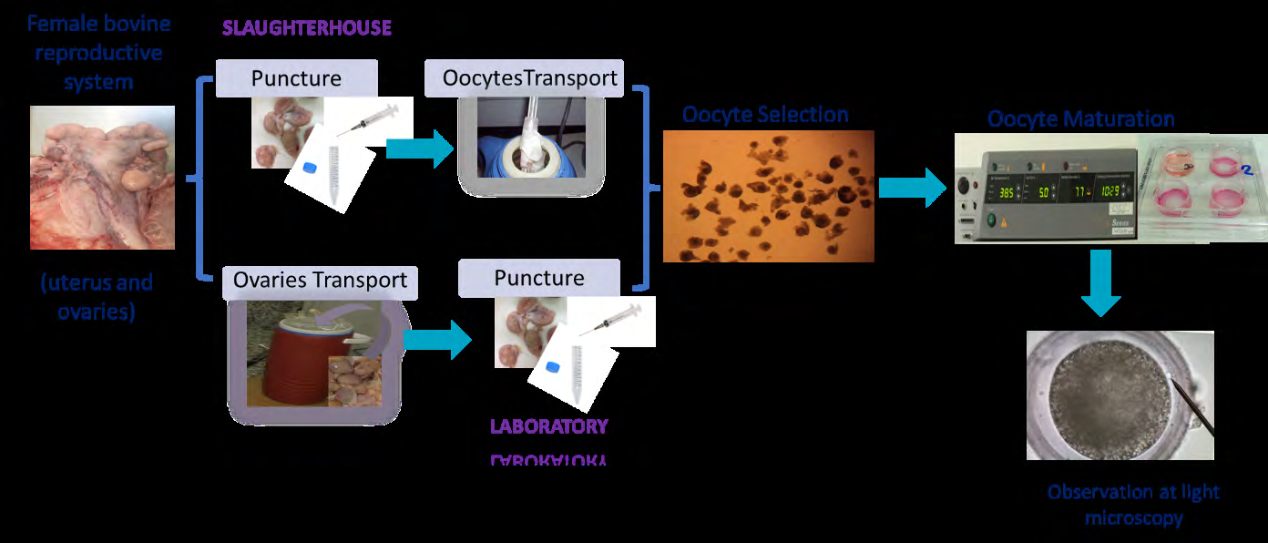
Puncture of bovine ovaries for oocyte collection at the slaughterhouse: The ovaries collected from cows at the slaughterhouse to be punctured in place, were immediately submerged in a Phosphate Buffer Saline 1x solution [PBS: 1.37M NaCl (Fisher Scientific, United States of America, USA), 27mM KCl (ChemLab, Belgium), 100mM Na2HPO4 (Fisher Scientific, New Hampshire, USA), 20mM KH2PO4 (ChemLab, Belgium)], supplemented with antibiotics (penicillin 100 IU/ml; streptomycin 100 µL/mL; Sigma-Aldrich, USA) and kept in a thermos bottle at 34-35°C. The complex-cumulus- oocytes (COCs) were aspirated from 3-8 mm follicles, with a 18G needle (Braun, Germany) attached to a 10 mL syringe (Terumo, Japan) and placed in a 50 mL falcon tube (Labbox, Spain) with oocyte collection medium. After collection, the fluid was poured into a 15 mL falcon tube (Labbox, Spain) containing maturation medium and transported to the laboratory in a thermos with water at 32ºC at room temperature (25-30°C). Upon arrival at the laboratory, 2 hours after puncture, the contents of the falcon tube were placed in a petri dish (90mm, VWR, USA). Puncture of bovine ovaries for oocyte collection at the laboratory: Those ovaries which were not processed immediately were collected from the same cows at the slaughterhouse and transported to the laboratory in a thermos at 32ºC at room temperature (25-30°C) in a container with PBS 1x solution supplemented with antibiotics. Once in the laboratory, the ovaries were washed in with PBS and kept in a water bath (J.P. Selecta, Spain) at 34-352°C. The puncture of ovaries and oocyte collection was performed as above described.
Bovine in Vitro Maturation of Oocytes (bIVM)
Culture media and solutions: The solutions used were the following: PBS solution 1x with antibiotics; collection medium: TCM 199 (Biochrom AG, United Kingdom), supplemented with HEPES (25mM, Fisher Scientific, USA), polyvinilic alcohol (PVA) (1mg/mL, Sigma-Aldrich, USA), antibiotics (penicilin 100 IU/mL; streptomycin 100 µL/mL, Sigma-Aldrich, USA), heparin (10 μg/mL, Sigma-Aldrich, USA) and glutamine (0.1 mg/mL, Alfa Aesar, USA); maturation medium: TCM 199, supplemented with Epidermal Growth Factor (10ng/ml, EGF, Gibco InVitrogen, USA), FSH/ LH (0.05 IU, Menopur, Ferring, USA), glutamine (0.1 mg/mL), sodium pyruvate (0.11 mg/ml, Sigma-Aldrich, USA) and PVA (1 mg/ mL). Experimental procedure: The experimental procedure used was the one described in ECVAM DB-ALM Protocol nº 129 -Toxicity test on in vitro maturation of bovine oocytes. Selection of oocytes and maturation: By using a stereo microscope (SZ, Olympus, Europe), grade I, II and III oocytes were selected, washed twice in collection medium, and transferred to a 4-wells tissue culture plate (VWR, USA) containing maturation medium (maximum 20 oocytes per well). This selection took place 8 hours after slaughter, and the time spent per group (oocytes punctured in the slaughterhouse and punctured in the laboratory) was about 30 minutes. The 4-wells plates were incubated at 5% CO2, 38.5°C, and saturated humidity for 24h in an incubator (UniTherm, CO2 Series, Uniequip GmbH, Germany). Oocyte denudation and microscopic examination: At the end of the incubation time, the granulosa and most of the cumulus oocytes cells were enzymatically removed, leaving only the corona radiata. For this procedure the oocytes of each well were collected and placed in contact with 100 µL of cumulase (Cumulase, Origio®, Denmark), for 1 minute and 20 seconds and then washed 5 times with maturation medium. The oocytes, after being stripped and placed in maturation medium, were observed under an inverted microscope (IX51, Olympus, Europe) under 400x magnification. Evaluation of oocytes maturation: The presence of a smaller, more compact refringent mass accompanying the secondary oocyte chromosomes corresponding to the extrusion of the first polar body (PB) associated to the second metaphase plate, was indicative of complete maturation. The number of oocytes in which PB extrusion was observed, and those in which it was not observed were registered. Oocytes in the germinal vesicle (GV), chromatin condensation (CC I and CC II) or germ vesicle disintegration (GVBD), metaphase I, anaphase I and telophase I stages were classified as non- matured [11].
The results were expressed as frequency of maturation (fm, %), considering the number of matured oocytes (mo) in Day Age (months) Animals (n) Frequency of matured oocytes (%; n) p-value Puncture at slaughterhouse Puncture at laboratory $$ 1 < = 1 3 \mathrm {m} 8 8 4 (2 7 / 3 2) 8 0 (2 8 / 3 5) \mathrm {N S} $$
1 >13m 7 87 (20/23) 77 (23/30) NS
$$ 2 < = 1 3 \mathrm {m} 9 9 5 (1 9 / 2 0) 8 5 (2 2 / 2 6) 0, 0 3 1 7 $$
2 >13m 9 100 (21/21) 94 (30/32) 0,0289
$$ 3 < = 1 3 \mathrm {m} 6 7 9 (3 3 / 4 2) 8 3 (3 5 / 4 2) \mathrm {N S} $$
3 >13m 5 85 (22/26) 90 (36/40) 0,0078
MEAN ± SD
$$ 8 8 \pm 8 \quad 8 5 \pm 6 \quad N S $$
NS: non-significant (p>0.05); SD: standard deviation. Table 2: Frequency of matured oocytes, after puncture at the slaughterhouse or at the laboratory. Number of matured oocytes in the total of punctured oocytes is also showed. The corresponding p-value highlighting statistical significance is showed, as well as data obtained for each group of animals.
We also analysed the results regarding the age of the animal at time of slaughter in Figure 2. Previous studies Morin L [12] have indicated that the age of the animal at time of puncture could influence the results. Specifically, oocytes the total of punctured oocytes (po):
Statistical Analysis
The differences between proportions (frequencies of maturation) were assessed for their statistical significance with the two-sided Chi-square test, using GraphPad prism v7.03 software (GraphPad Software, San Diego, California, USA). Results were statistically significant when p-value <0.05.
Results and Discussion
This study was designed to investigate if there were differences regarding maturation of oocytes obtained by puncture of ovaries at the slaughterhouse in comparison to those punctured at the laboratory.
The maturation rate of the oocytes collected from ovaries punctured at the slaughterhouse was higher (88±8%), when compared with the one obtained when the ovaries were punctured at the laboratory (85±6%), indicating that transport time and/or conditions does not influence maturation rates in Table 2. We can refer that our mean maturation (86 ± 7%) (mean±SD) is corroborated by other studies. Therefore, the differences found could be possibly attributed to biological (individual) variance and not to puncture site interference. In fact, and supporting this hypothesis, we found statistically significant differences when considering the average maturation rates determined in our study by grouping the animals in two different age groups in Table 2.
collected from younger animals (pre-pubertal calves) have been shown to be less competent. In fact, when grouping the animals by age group we did find some statistical significance in Table 2, meaning that the age of the animal at time of puncture, and particularly considering the 13 months categorisation, could influence oocyte competence. There was also no statistically significant difference regarding day of puncture in Figure 3.
![Figure 2: Previous studies Morin L [12] have indicated that the age of the animal at time of puncture could influence the results. Specifically, oocytes the total of punctured oocytes (po):](/fulltextimages/9568/fig_2.jpeg)
To the best of our knowledge, previous studies only report collection of ovaries at the slaughterhouse, and their puncture at the laboratory following transport. Of these, studies have reported maturation frequencies of 30-40%. Our results regarding frequency of maturation of oocytes punctured at the laboratory, after transport, are higher:
puncture at the slaughterhouse (88±8%), puncture at the laboratory (85±6%). The lower maturation rates obtained in the literature could be explained by several factors, namely environmental factors, such as the time between puncture and maturation and the removal of the oocyte from its follicle environment itself, which depending on the developmental stage could have molecular and hormonal negative impact [13]. Other studies, that report ovum pick- up (OPU) procedures, in which oocytes are directly obtained from live cows, report cleavage (after blastocyst formation) frequencies higher than 60% [14, 15, 16]. In this case, maturation rates are expected to be higher, since viability and hormonal state of the oocytes are more easily maintained [17]. However, that technique is expensive, needs a lot of specialized equipment’s (ultrasound, transvaginal probe, suction pump and guide) and highly skilled personnel with technical knowledge [18, 19, 20, 21]. By using discarded material collected at slaughterhouses, the oocyte bIVM test is fully in accordance with the 3R’s principle (reduction, re-use, recycle) for animal testing. However, this methodology is limited to a 4-hours timeframe after collection of the ovaries. Therefore, in our study, we consider this 4-hour timeframe to understand if puncture of collected ovaries in a laboratory after transport would be feasible. We obtained very high maturation rates (>80%), considering that the maturation rate of oocytes collected at slaughterhouses usually never reaches more than 40% [7]. Therefore, the 4-hour timeframe is validated by our study, which strengthens the use of slaughterhouse material for this technique.
Conclusion
Considering the average maturation rates obtained, and the lack of overall statistically significant differences between the two methodological approaches, we conclude that for implementation of oocyte bIVM tests transport of the ovaries and puncture in laboratory is a better option, since it is a more clean, convenient and controllable procedure.
Conflict of Interest
The authors declare having no conflict of interest.
Declaration of Funding
This work was supported by ”INOVEP project – Innovation with Plant Extracts”, I&DT projects for companies in collaboration with scientific entities, project number 33815, Centro2020.
Data Availability Statement
This study is a work of review. All references cited are available in databases used for the search.
References
-
Lonergan P, Fair T (2016) Maturation of Oocytes in Vitro. Annual Review of Animal Biosciences 4: 255-268.
-
Farsi MM, Kamali N, Pourghasem M (2013) Embryological aspects of oocyte in vitro maturation. International Journal of Molecular and Cellular Medicine 2: 99-109.
-
Blanco M, Demyda S, Moreno Millán M, Genero E (2011) Developmental competence of in vivo and in vitro matured oocytes: A review. Biotechnology and Molecular Biology Reviews 6: 155-165.
-
Sirard MA, Blondin P (1996) Oocyte maturation and IVF in cattle. Animal Reproduction Science 42(1-4): 417- 426.
-
European Centre for the validation of Alternative Methods (ECVAM) (1996) DB-ALM Protocol n° 129: Toxicity Test on In Vitro Maturation of Bovine Oocytes. pp: 1-14.
-
Beker Van Woudenberg A, Gröllers-Mulderij M, Snel C, Jeurissen N, Stierum R, et al. (2012) The bovine oocyte _in_ _vitro_ maturation model: a potential tool for reproductive toxicology screening. Reproductive Toxicology 34(2): 251-260.
-
Lonergan P, Fair T (2014) The ART of studying early embryo development: Progress and challenges in ruminant embryo culture. Theriogenology 81: 49-55.
-
Mermillod P, Dalbiès Tran R, Uzbekova S, Thélie A, Traverso JM, et al. (2008) Factors affecting oocyte quality: who is driving the follicle? Reproduction in Domestic Animals 43(S2): 393-400.
-
Park YS, Kim SS, Kim JM, Park HD, Byun MD (2005) The effects of duration of in vitro maturation of bovine oocytes on subsequent development, quality and transfer of embryos. Theriogenology 64: 123-134.
-
Penitente Filho JM, Jimenez CR, Zolini AM, Carrascal E, Azevedo JL, et al. (2015) Influence of corpus luteum and ovarian volume on the number and quality of bovine oocytes. Animal Science Journal 86(2): 148-152.
-
Smiljakovic T, Tomek W (2006) Meiotic maturation and in vitro maturation of bovine oocytes. Biotechnology in Animal Husbandry 22: 29-34.
-
Morin-Doré L, Blondin P, Vigneault C, Grand FX, Labrecque R, et al. (2020) DNA methylation status of bovine blastocysts obtained from peripubertal oocyte donors. Molecular Reproduction and Development 87(8): 910-924.
-
Gottardi FP, Mingoti GZ (2010) Bovine oocyte maturation and influence on subsequent embryonic developmental competence. Revista Brasileira de Reprodução Animal 33(2): 82-94.
-
Boland MP, Lonergan P, O Callaghan D (2001) Effect of nutrition on endocrine parameters, ovarian physiology, and oocyte and embryo development. Theriogenology 55(6): 1323-1340.
-
Fouladi-Nashta A, Gutierrez CG, Gong JG, Garnsworthy PC, Webb R (2007) Impact of dietary fatty acids on oocyte quality and development in lactating dairy cows. Biology of Reproduction 77: 9-17.
-
Galli C, Crotti G, Notari C, Turini P, Duchi R, et al. (2001) Embryo production by ovum pick up from live donors. Theriogenology 55(6): 1341-1357.
-
Merton JS, de Roos APW, Mullaart E, de Ruigh L, Kaal L, et al. (2003) Factors affecting oocyte quality and quantity in commercial application of embryo technologies in the cattle breeding industry. Theriogenology 59(2): 651- 174.
-
Ferré LB, Kjelland ME, Strøbech LB, Hyttel P, Mermillod P, et al. (2020) Review: Recent advances in bovine in vitro embryo production: reproductive biotechnology history and methods. Animal 14(5): 991-1004.
-
Hagemann LJ, Beaumont SE, Berg M, Donnison MJ, Ledgard A, et al. (1999) Development during single IVP of bovine oocytes from dissected follicles: Interactive effects of estrous cycle stage, follicle size and atresia. Molecular Reproduction and Development 53(4): 451- 458.
-
Luciano AM, Sirard MA (2018) Successful in vitro maturation of oocytes: a matter of follicular differentiation. Biology of Reproduction 98(2): 162-169.
-
Quezada Casasola A, Eugenia Martínez Armendáriz K, Fabián Itzá-Ortiz M, María Escárcega-Ávila A, Pérez- Eguía E, et al. (2018) Effect of presence of corpora lutea on cumulus expansion of in vitro matured bovine oocytes selected by trypan blue and brilliant cresyl blue tests. Journal of Applied Animal Research 46: 967-972.
- California Red-Legged Frog and Non-Listed Amphibians Response to Non-Native Fish Removal
- Industrial Standardization of the Bio-OS: Algorithmic Codification of Resilience Engineering Guidelines and Version V8 Architecture
- Climate Variability and the Sustainability of Snail Farming in Nigeria: Past Trends, Present Challenges and Potential Outlook
- The Evaluation of the Surveillance System of Anthrax in Gilgit-Baltistan, Pakistan, 2018
- Natural Decline to Extinction of A New Zealand Rabbit Population
- Mitochondrial Bio-Logistics: Steering Co-Enzyme Q10 and Lycopene Synergies within the Science 4.0 Bio-OS Framework