A Case of Negligence in the Diagnosis of Canine Juvenile Demodicosis. Case Report
Canine demodicosis is a parasitic dermatopathy caused by Demodex canis, a mite that is part of cutaneous microbiota of dogs. Its overgrowth inside hair follicles and sebaceous glands causes alopecia and erythema in the localized form, and its generalized form usually goes along with a severe case of folliculitis. Its diagnosis is performed through skin scraping, trichogram by epilation or impression with acetate tape of the affected areas and visualization of material by optical microscopy. The treatment consists by the use of specific parasiticides, and in cases of opportunistic bacterial infection, the use of antibiotics is recommended. This case report describes the generalized form of demodicosis in a 2-month-old male puppy, Dachshund breed, whose diagnosis was initially was not defined by not performing a skin scraping and viewing the material obtained under a microscope.
Introduction
The Demodex canis is a mite that is part of the skin microbiota, present in small amounts inside of hair follicles and sebaceous glands of the skin of healthy dogs. The transmission of mite occurs from the mother to the newborn infants through direct contact in the first two or three days of neonatal life [1].
In some dogs, there is an excessive proliferation of mites inside the hair follicles, leading to the emergence of canine demodicosis, a non-contagious parasitic dermatopathy. The exacerbated proliferation of D. canis results in an inflammatory dermatosis that can be classified as localized demodicosis or generalized demodicosis and, depending on the first clinical manifestations it can be classified as presenting a juvenile or adult character [2].
Localized demodicosis usually presents itself as a condition with a benign course, with spontaneous resolution in most cases. Animals under one year of age are most often affected. The lesions are characterized by alopecic and erythematous areas, usually located in cephalic region and/ or forelimbs. The generalized demodicosis is considered a serious dermatopathy in dogs, where the lesions present in the form of severe folliculitis with hemorrhagic exudation accompanied by opportunistic bacterial infection [2, 3].
The diagnosis is make through deep skin scraping, and the trichogram can also be performed by epilation or printing with acetate tape in the periocular, perilabial or interdigital regions [4, 5, 6].
Case Report
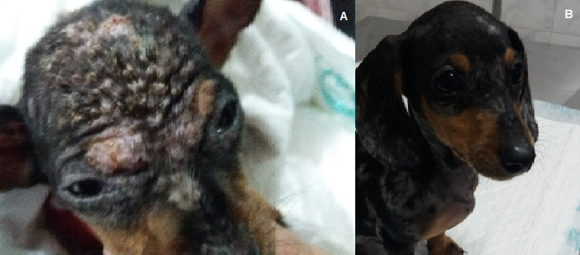
In June 2022, an animal of the canine species, Dachshund breed, 02 months old, male, weighing 1.0 kg, was attended, where the tutor reported the presence of erythematous crusted areas and pustules with purulent secretion on the head (Figura 1A) and body. According with the tutor, the animal was taken to a colleague who underwent a clinical examination and stated that the wounds were the result of licking carried out by another animal belonging to the tutor. No additional examination was performed, and ointment based on dexamethasone+neomycin sulfate+ griseofulvin+benzocaine was prescribed. On clinical examination, the patient was not very active, the mucous membranes were normally colored and the submandibular and popliteal lymph nodes were slightly enlarged. He had a body temperature of 390C, normal skin tone and absence of reaction to abdominal palpation. A blood sample was collected for a complete blood count and biochemical examination.
A skin scraping was performed using a sterile scalpel blade in three different areas of the body, where the material was placed on a glass slide, clarified with 10% KOH, coverslipped and visualized under optical microscopy at 10x and 40x magnification. A large number of mites were observed per field, confirming the diagnosis of generalized demodicosis.
The patient was medicated with cephalexin (25mg/ kg) orally every 12 hours for 14 days and ivermectin 0.4mg/kg orally for 30 days. The hemogram showed mild lymphopenia (typical lymphocytes=1033/mm3 of blood; normal values=1500 to 7000/mm3 of blood). In the other exams, the parameters were within the normal limits for the age and species. The skin scraping was repeated on the thirtieth day of treatment with a negative result. After 60 days of treatment, the animal was in good clinical condition (Figure 1B) and new scrapings were performed, where no mites were observed.
Discussion
The mites of the gender Demodex spp. are commensal parasites found in hair follicles and sebaceous glands in most animal species. There are three known species of mites in the gender Demodex on dogs: Demodex canis, Demodex injai e Demodex cornei [7].
The demodectic scabies is a chronic pathology that occurs when there is excessive proliferation of the D. canis mite inside the hair follicles and sebaceous glands of dogs [8].
The affected animals may have the localized or generalized form, which is classified according to age as juvenile (below 18 months of age) or adult. The generalized form is considered more severe, being accompanied by exuding pustules and scabs [3].
In this case report, performing a parasitological examination of the skin scraping was essential for the diagnosis, a fact that was ignored in the first consultation of dog. The first technique of choice for diagnosing demodicosis is the parasitological examination of skin scrapings, which is easy to perform, low cost and highly sensitive [6]. The skin scrapings must be deep and carried out in the direction of hair growth, performing in different regions of the body, especially in areas of transition from healthy skin to the lesion and with the presence of comedones. In animals with multifocal lesions or generalization of demodicosis, it is recommended to examine skin scrapings from three to six sites [6, 9]. The diagnosis is made when a large number of adult mites are observed or when there are large number of eggs, larvae or nymphs [6].
The blood count of the animal in this case report revealed a mild lymphopenia. In young animals with generalized demodicosis, the literature describes a decrease in the number of circulating T lymphocytes [10], possibly related with the process of premature apoptosis of this cell type [11], in addition to increased serum levels of interleukin-10 (IL- 10) and cholinesterase activity; decreased levels of tumor necrosis factor alpha (TNF-α) [12]; and upregulation of Toll like receptor 2 (TLR2) and downregulation of TLR4 and TLR 6 [13, 14], factors associated with immunosuppression that favors the exacerbated growth of mites.
For the diagnosis of demodicosis, it is important to carry out deep scrapings on the skin, trichogram or impression with acetate tape, what was not done in the first consultation. In the rare cases a skin biopsy may be required for the diagnosis [5, 6, 15].
Because the animal had an opportunistic bacterial infection, the use of cephalexin was prescribed, being essential the use of antibiotics such as cephalexin, enrofloxacin or amoxicillin+clavulanic acid in the treatment of secondary bacterial infection [16, 17, 18].
The treatment option for generalized demodicosis was decided to use ivermectin due to its easy availability and low cost in Brazil. The literature successfully reports the use of parasiticide drugs such as ivermectin [19, 20], moxidectin and imidacloprid [21], isoxazolines [22] or fluralaner [23, 24] to control of mite population.
Conclusion
In this case report highlights the importance of performing skin scrapings in all cases of cutaneous dermatopathies for the diagnosis of demodicosis and its correct treatment.
References
-
Greve JH, Gaafar SM (1966) Natural transmission of Demodex canis in dogs. J Am Vet Med Assoc 148(9): 1043-1045.
-
Sing SK, Dimri U (2014) The immuno-pathological conversions of canine demodicosis. Vet Parasitol 203(1- 12): 1-5.
-
Kelly PA, McKay JS, Maguire D, Jones M, Roberts, et al. (2022) A retrospective study of cases of canine demodicosis submitted to a comercial diagnostic laboratory servicing the United Kingdom and Ireland (2017-2016): Part 1-Signalment, lesions distribution, treatment, and concurrent disease. Res Vet Sci 30(153): 99-104.
-
Fondati A, De Lucia M, Furiani N, Monaco M, Ordeix L, et al. (2010) Prevalence of Demodex canis-positive healthy dogs at trichoscopic examination. Vet Dermatol 21(2): 146-151.
-
Barillas OF, Bajwa J, Guillot J, Arcique A (2019) Comparison of acetate tape impression, deep skin scraping, and microscopic examination of hair for therapeutic monitoring of dogs with juvenile generalized demodicosis: a pilot study. Can Vet J 60(6): 596-600.
-
Mueller RS, Rosenkrantz W, Bensignor E, Karas-Tecza J, Paterson T, et al. (2020) Diagnosis and treatment of demodicosis in dogs and cats: clinical consensus guidelines of the World Association for Veterinary Dermatology. Vet Dermatol 31(1): 5-27.
-
Hu L, Zhao Y, Zhang W (2022) De novo transcriptome sequencing and functional annotation of Demodex canis. Exp Appl Acarol 87(2-3): 219-233.
-
Paichitrojjana A (2022) Demodex: the worst enemies are the ones that used to be friends. Dermatol Reports 14(3): 9339.
-
Pereira AV, Pereira AS, Gremião ID, Campos MP, Ferreira AM (2012) Comparison of acetate tape impression with squeezing versus skin scraping for the diagnosis of canine demodicosis. Aust Vet J 90(11): 448-450.
-
Oliveira CD, Larsson CE, de Camargo MM (2015) Longitudinal assessment of T-lymphocyte subpopulations during generalized demodicosis in dogs and their relationship with remission. Vet Dermatol 26(1): 18-22.
-
Singh SK, Dimri U, Sharma MC, Swarup D, Sharma B, et al. (2011) The role of apoptosis in immunosuppression of dogs with demodicosis. Vet Immunol Immunopathol 144(3-4): 487-492.
-
Kumari P, Nigam R, Singh A, Nakade UP, Sharma A, et al. (2017) Demodex canis regulates cholinergic system mediated immunossupressive pathways in canine demodicosis. Parasitology 144(10): 1412-1416.
-
Kumari P, Nigam R, Choudhury S, Singh SK, Yadav B, et al. (2018) Demodex canis targets TLRs to evade host immunity and induce canine demodicosis. Parasite Immunol 40(3): 1-15.
-
Soman SP, Singh SK, Kumari P, Chodhury S, Singh A, et al. (2020) Quantification of Immuno-regulatory cytokine and Toll-like receptors gene expression. in dogs with generalized demodicosis. Vet Parasitol 280: 109063.
-
Mueller RS, Bensignor E, Ferrer L, Holm B, Lemarie S, et al. (2012) Treatment of demodicosis in dogs: 2011 clinical practice guidelines. Vet Dermatol 23(2): 86-96.
-
Kuznetsova E, Bettenay S, Nikolaeva L, Majzoub M, Mueller R (2012) Influence of systemic antibiotics on the treatment of dogs with generalized demodicosis. Vet Parasitol 188(1-2): 148-155.
-
Jacobs S, Van Daele MA, Brown JN (2019) Treatment of Demodex-associated inflammatory skin condition: a systematic review. Dermatol Ther 32(6): e13103.
-
Kelly PA, McKay JS, Maguire D, Jones M, Roberts L, et al. (2022) A retrospective study of cases of canine demodicosis submitted to a comercial diagnostic laboratory servicing the United Kingdom and Ireland (2017-2018) Part 2; aerobic culture and antimicrobial susceptibility results. Res Vet Sci 153: 92-98.
-
Rahamn M, Bostami MB, Datta A, Al Momen Sabuj A, Rana EA, et al. (2021) Estimation of the prevalence and determination of risk factors associated with demodicosis in dogs. J Adv Vet Anim Res 8(1): 116-112.
-
Li J, Wei E, Reisinger A, French LE, Clanner-Engleshofen BM, et al. (2023) Comparison of different anti-Demodex strategies: a systematic review and meta-analysis. Dermatology 239(1): 12-31.
-
Paterson TE, Halliwell RE, Fields PJ, Louw ML, Ball G, et al. (2014) Canine generalized demodicosis treated with varying doses of a 2.5% moxidectin+10% imidacloprod spot-on and oral ivermectin: parasiticidal effects and long-term treatment outcome. Vet Parasitol 205(3-4): 687-696.
-
Defalque VE (2022) Isoxazolines for treating canine demodicosis, sarcoptic mange (scabies), and lice infestation. Can Vet J 63(11): 1159-1162.
-
Peterson I, Chiummo R, Zschiesche E, Karas-Tecza J, Rapti D, et al. (2020) A European field assessment of the efficacy of fluralaner (Bravecto®) chewable and spot- on formulations for treatment of dogs with generalized demodicosis. Parasit Vectors 13(1): 304.
-
Rohdich N, Meyer L, Guerino F (2022) Fluralaner 5.46% (w/w) flavored chewable tablet (Bravecto® 1-month) is effective for treatment of canine generalized demodicosis. Parasit Vectors 15(1): 83.
- California Red-Legged Frog and Non-Listed Amphibians Response to Non-Native Fish Removal
- Industrial Standardization of the Bio-OS: Algorithmic Codification of Resilience Engineering Guidelines and Version V8 Architecture
- Climate Variability and the Sustainability of Snail Farming in Nigeria: Past Trends, Present Challenges and Potential Outlook
- The Evaluation of the Surveillance System of Anthrax in Gilgit-Baltistan, Pakistan, 2018
- Natural Decline to Extinction of A New Zealand Rabbit Population
- Mitochondrial Bio-Logistics: Steering Co-Enzyme Q10 and Lycopene Synergies within the Science 4.0 Bio-OS Framework